| ABSTRACTS P-357 to P-536 POSTER PRESENTATIONS
Posters will be on display throughout the symposium, but will be attended by their presenting authors as follows: Odd numbers on Friday 11:00 - 12:00, Sunday 11:00 - 12:00
and Monday 10:00 - 11:00 |
||||||||||||||||||||||||||||||||||||||||||||||||||||||||||||||||||||||||||||||||||||||||||||||||||||||||||||||||||||||||||||||||||||||||||||||||||||||||||||||||||||||||||||||||||||||
Osteoporosis: Pathophysiology, Genetics, Epidemiology Continued...
EFFECT OF FLUOROURACIL ON BONE REMODELING IN RATS U. Cegiela*, W. Janiec, J. Folwarczna, I. Kaczmarczyk-Sedlak, M. Pytlik, B. Nowińska, L. Sliwiński Department of Pharmacology, Medical University of Silesia, Sosnowiec, Poland Intensification of bone resorption is often observed in neoplastic diseases. Several factors released by tumor cells, or by immune cells responding to tumor cells, are known to enhance osteoclastic activity. Chemotherapy, associated with the risk of damaging effect on normal, especially rapidly-dividing cells, may also contribute to disorders of bone remodeling. The aim of the present study was to examine the effect of fluorouracil (5-FU) on the macrometric and histomorphometric parameters of bones, as well as the mechanical properties of the femur. The experiments were carried out on 24 Wistar rats (11-week-old), divided into 4 groups: I Control, II FU-30 (5-FU: 30 mg/kg p.o. daily for 5 days every 2 weeks), III FU-15 (5-FU: 15 mg/kg i.m. daily for 5 days every 2 weeks), IV FU-65 (5-FU: 65 mg/kg i.m. once weekly). The animals were sacrificed after 4 weeks of the experiment. Fluorouracil disturbed bone remodeling in rats, inducing decreases in the width of trabeculae in the epiphysis and metaphysis of the femur, width of periosteal and endosteal osteoid, periosteal and endosteal transverse growth, and area of the transverse cross-section in the tibia. The calcium and mineral content in bones also decreased. The observed disorders of bone formation and mineralization, caused by 5-FU, led to worsening of mechanical properties of the femur (decreases in the load necessary to cause the fracture of the diaphysis and the neck of the femur). ASSOCIATION BETWEEN POLYMORPHISMS OF THE TCIRG1 GENE AND BONE MASS IN PERIMENOPAUSAL WOMEN C. Sobacchi1*, P. Vezzoni1, D. M. Reid2, F. E. A. McGuigan2, M. Mirolo1, A. Frattini1, O. M. E. Albagha2, A. Musio1, A. Villa1, S. H. Ralston2 1Istituto Tecnologie Biomediche, CNR, Milan, Italy 2Department of Medicine and Therapeutics, University of Aberdeen, Aberdeen, UK The TCIRG1 gene on chromosome 11q12-13 encodes a component of the osteoclast vacuolar proton pump. Previous work has shown that autosomal recessive osteopetrosis is due to inactivating mutations of the TCIRG1 gene in about 50% of cases. In order to investigate whether allelic variation in TCIRG1 contributes to the regulation of bone mass in normal individuals, we studied the relationship between bone mineral density (BMD) and polymorphisms of the TCIRG1 gene in a population based cohort of 739 perimenopausal women. Mutation screening of the promoter, exons and intron-exon boundaries identified five polymorphisms of TCIRG1; two in the promoter (G-1082A and G-900A); two within intron 4 (C3658T and A3900G) and one within intron 11 (G8645A). All polymorphisms were in strong linkage disequilibrium (LD) with the exception of C3856T where significant LD was observed only with A3900G. A nalysis of the genotype data using the Haplotyper program predicted 31 different haplotypes in the study population, although five common haplotypes accounted for 77.3% of alleles found in our population. There was a significant association between the G-1082A polymorphism and BMD at the lumbar spine (p=0.01) and femoral neck (p=0.042), with lower BMD values in carriers of the -1082A allele. This association remained after correcting for age, weight, menopausal status, smoking and HRT use (p=0.008 for spine BMD and p=0.03 for hip BMD). None of the other polymorphisms or common haplotypes were significantly associated with BMD. We conclude that, in this relatively large population, allelic variation at the G- 1082A polymorphic site at the TCIRG1 locus accounts for part of the heritable component of BMD, raising the possibility that TCIRG1 alleles act as a genetic determinant of bone mass in Scottish women. ROLE OF DIETARY CALCIUM INTAKE IN INFLUENCING BODY WEIGHT, BONE MASS AND PREVALENCE OF OSTEOPOROSIS IN EARLY POSTMENOPAUSAL WOMEN M. Varenna, L. Binelli, F. Zucchi, S. Casari*, L. Sinigaglia Department and Chair of Rheumatology, University of Milan, 'G. Pini' Institute, Milan, Italy Several studies have shown that a high level of calcium intake reduces bone loss in the appendicular skeleton of late postmenopausal women, but it seems to be ineffective in stopping vertebral bone loss in early postmenopausal women. However, this variable could be underestimated on the light of the results of epidemiological and biochemical studies about the inverse relationship between calcium intake and body weight, and overweight could play a protective role on bone mass in women with low calcium intake. To verify this hypothesis cases were recruited among women who were referred to our hospital for their first lumbar bone mineral density (BMD) measurement (Hologic QDR 4500) and had undergone spontaneous menopause five or fewer years earlier. Quantitative estimates of calcium intake were obtained from a 7-day food frequency questionnaire. By using calcium intake quartiles, we selected 465 women (age: 53.9 ±SD 2.9; age at menopause: 51.3 ±SD 2.2) in the upper quartile (calcium intake more than 750 mg/day), and 426 women (age: 53.7 ±SD 2.7; age at menopause: 50.9 ±SD 2.1) in the lower quartile (calcium intake less than 400 mg/day). No difference in age and age at menopause was found between the groups. As found in other epidemiological studies, women with lower calcium intake showed a greater Body Mass Index (BMI) and a higher prevalence of overweight (respectively 24.1 ±SD 3.4 vs 23.2 ±SD 3.3; p<0.001 and 37.3% vs 13.3%; p<0.001). Lumbar BMD was slightly lower in women with low calcium intake (0.86 ±SD 0.12 vs 0.89 ±SD 0.12), but no difference was found in Osteoporosis (OP) prevalence (20.9% vs 17.6%; p=0.2). Nevertheless, when BMI was entered in the analyses, both a generalized linear model for BMD value and a stepwise multiple logistic regression analysis for OP risk showed that calcium intake significantly influenced BMD values (p<0.001) and acted as significant predictive factor for OP risk (OR 1.38, 95% CI 1.03-1.85 for women in the lower quartile). In conclusion, the effective value of calcium intake in preventing bone loss in early postmenopausal women could be overshadowed by the protective role exerts by a greater body weight in women with low calcium intake. EFFECT OF CONCURRENT ADMINISTRATION OF ALENDRONATE SODIUM AND RETINOL ON THE MECHANICAL PROPERTIES OF THE FEMUR IN OVARIECTOMIZED RATS L. Sliwiński*, M. Pytlik, I. Kaczmarczyk-Sedlak, W. Janiec, J. Folwarczna, B. Nowińska, U. Cegiela, W. Pytlik Department of Pharmacology, Medical University of Silesia, Sosnowiec, Poland Alendronate sodium, an aminobisphosphonate with potent antiresorptive activity, is used in the treatment of postmenopausal osteoporosis. Retinol, as a component of multivitamin preparations, is frequently used especially by elderly people. There are no reports on the interaction of alendronate sodium and retinol. The aim of the present study was to investigate the effect of administration of alendronate sodium and retinol on mechanical properties of the femoral bone in bilaterally ovariectomized rats. The experiments were carried out on 3-month-old Wistar rats, divided into 7 groups: I - (C) sham-operated control rats, II - (OVX) ovariectomized control rats, III - (OVX + ALN) ovariectomy + alendronate sodium 3 mg/kg p.o., IV - (OVX + K700) ovariectomy + retinol 700 IU/kg p.o., V - (OVX + R3500) ovariectomy + retinol 3500 IU/kg p.o., VI - (OVX + ALN + R700) ovariectomy + alendronate sodium 3 mg/kg p.o. + retinol 700 IU/kg p.o., VII (OVX + ALN + R3500) ovariectomy + alendronate sodium 3 mg/kg p.o. + retinol 3500 IU/kg p.o. The drugs were administered to the rats by daily oral gavage (alendronate sodium in the morning, retinol in the afternoon) for 28 days. The mechanical properties of the diaphysis and neck of the femur, body mass gain, bone mass, bone mineral content and calcium content (in the tibia, femur and L-4 vertebra) were examined. Bilateral ovariectomy induced osteopenic changes in the rat skeletal system. Alendronate sodium (3 mg/kg p.o.) counteracted the development of osteopenia induced by ovariectomy. Retinol in both used doses unfavourably affected the examined bone parameters of ovariectomized rats. Retinol administered with alendronate sodium lessened the preventive action of alendronate sodium on the development of osteopenic changes in the skeletal system of ovariectomized rats. OESTROGEN AND THE CLUSTERING OF CORTICAL REMODELLING N. Loveridge1*, S. Vedi2, K. L. Bell1, J. E. Compston2, J. Reeve1 1Bone Research Group (MRC), Dept of Medicine, Addenbrookes Hospital (Box 157), Cambridge, UK 2Bone Research Group, Dept of Medicine, Addenbrookes Hospital (Box 157), Cambridge, UK Femoral neck fracture is strongly associated with the rapid appearance of large cortical pores. These pores are related to the spatial clustering of osteonal remodelling activity as identified by the presence of osteoid on the canal surface. However, the cause of this clustering remains an enigma. To determine whether oestrogen regulates clustering we have analysed iliac biopsies from post-menopausal women before and after 2 years treatment with HRT (n=10) and from pre-menopausal women before and after 6 months treatment with GnRH (n=10) for the presence of clustering of both resorbing and forming canals. For each biopsy the number and location of resorbing (crenellated) and forming (osteoid) canals were noted. Cluster analysis (JMP software) used 0.32mm (2x the mean inter-osteonal distance for all biopsies) as the cluster radius. For the HRT group, the percentage of resorbing and forming canals that were clustered was significantly higher than would be expected by chance (resorbing:- +22.2±7%. p=0.007; forming: +25±5%, p<0.0001). However, HRT treatment did not affect clustering of either resorbing or forming canals (Resorbing:- Pre 40.5±9%, Post 38.9±13% p=0.93; Forming:- Pre 49.9±7.5%, Post 57.7±7%, p=0.46). For the GnRH group there was significant clustering of both resorbing and forming canals (resorbing:- +14±6%. p=0.03; forming: +15±3%, p<0.0001;) but the inhibition of oestrogen secretion had no effect (Resorbing:- Pre 48.3±9%, Post 52±9% p=0.73; Forming:- Pre 53.1±7.4%, Post 55.1±7.5%, p=0.85.). Clustering was still significant at a radius of 0.16mm but was unaffected by treatment in either group. This study has shown for the first time, that in the ilium, a significant percentage of those canals undergoing resorption as well as those undergoing formation are spatially clustered. Neither the presence (HRT treatment) nor absence (GnRH treatment) of oestrogen affected the proportion of clustered canals. This suggests that oestrogen influences remodelling activity by regulating activation of complete clusters as single functional units rather than the individual canals of which clusters are composed. Therefore, estrogens prevent fractures by mechanisms that do not involve the prevention of clustering. Understanding how clustering is regulated may open the way to treatments for osteoporosis which augment the effectiveness of oestrogens and activators of oestrogen receptors. THE ANALYSIS OF ASSOCIATION BETWEEN ALLELES OF ERALPHA AND BGLAP GENES AND OSTEOPOROSIS IN POSTMENOPAUSAL WOMEN FROM RUSSIA M. V. Moskalenko*, M. V. Aseev, I. E. Zazerskaya Ott's Institute of Obstetrics and Gynecology RAMS, St-Petersburg, Russia Osteoporosis is a common multifactor disease with a strong genetic component. It is characterised by decreasing bone mineral density (BMD) and microarchitectural deterioration, which leads to increased susceptibility to fracture. Nowadays a number of genes have been identified, which play important role in regulation of Ca 2+ concentration and contribute in bone metabolism. For several genes exact mutations were discovered, which resulted in activity changes of corresponding proteins or in forming of functionally abnormal products. The allele of ERalpha and BGLAP (osteocalcin) genes in 69 non-related individuals without osteoporosis rates Northwest Russian population (control group) and in 40 postmenopausal women with osteoporosis (1 group) and 57 postmenopausal of women with severe form of osteoporosis (2 group) were investigated. The polymorphisms ERalpha and BGLAP genes were studied by PCR-RFLP method. The RFLPs were represented as X or x (XbaI) and P or p (PvuII) for ERalpha gene; as H or h (HindIII) for BGLAP gene, with the lower case letters signifying the presence of restriction site. We have not elucidated any authentic differences in frequencies of alleles of XbaI and PvuII polymorphism in 1 intron of ERalpha gene in group 1 of patients, compared to the group 2 and it's average frequency in population (p>0.05). The frequencies of alleles of this polymorphism were 25,6 % (X) and 38,9% (P) in control group; 38,7% (X) and 47,5% (P) in group 1; 35,1% (X) and 46,5% (P) in group 2. The frequency of functionally abnormal allele H (HindIII polymorphism of BGLAP gene) in group 2 was 23,7%, which is significantly higher (p<0,01) than only in group 1 (10,0%). The frequency of this allele in control group was 18,1% and did not significantly differ from the average in group 1 and group 2 (p>0,05). According to obtained data the ERalpha polymorphism did not demonstrate any major effect of on pathogenesis of osteoporosis of postmenopausal Russian women. The present study also suggests that the presence of the H allele of BGLAP gene is predictive factor for severe form of osteoporosis only. EFFECT OF ADMINISTRATION OF RETINOL AND ETIDRONATE ON BONE HISTOMORPHOMETRIC PARAMETERS IN OVARIECTOMIZED RATS I. Kaczmarczyk-Sedlak*, W. Janiec, M. Pytlik, L. Sliwiński, J. Folwarczna, U. Cegiela, B. Nowińska Department of Pharmacology, Medical University of Silesia, Sosnowiec, Poland Retinol belongs to factors affecting bone remodeling. The effect of retinol on the osseous tissue depends on the dose and duration of treatment. Retinol can cause bone damage and deformation. Retinol is frequently administered chronically in too high doses, sometimes by osteoporotic patients. The aim of the present study was to examine the interaction between retinol and an antiresorptive drug - etidronate disodium in bilaterally ovariectomized rats. The experiments were carried out on Wistar rats (200±30 g), divided into 7 groups: I - sham operated control rats, II - ovariectomized control rats (OVX), III - OVX + etidronate (10 mg/kg p.o.), IV - OVX + retinol (700 IU/kg p.o.), V - OVX + retinol (3500 IU/kg p.o.), VI - OVX + etidronate (10 mg/kg p.o.) + retinol (700 IU/kg p.o.), VII - OVX + etidronate (10 mg/kg p.o.) + retinol (3500 IU/kg p.o.). The drugs were administered for 4 weeks. Bone macrometric and histomorphometric parameters of the tibia (transverse growth, width of periosteal and endosteal osteoid, area of the transverse cross-section of the diaphysis and area of the transverse cross-section of the marrow cavity) and the femur (width of epiphyseal and metaphyseal trabeculae, width of epiphyseal cartilage) were examined. Etidronate partially counteracted the development of changes induced by the ovariectomy. Retinol (700 IU/kg p.o.) caused decreases in the area of the transverse cross-section of the marrow cavity and the width of osteoid, and an increase in the width of trabeculae. Retinol (3500 IU/kg p.o.) caused decreases in bone mass and the area of the transverse cross-section of the marrow cavity, and an increase in the width of trabeculae. Concurrent administration of etidronate and retinol in ovariectomized rats seemed not to affect bone histomorphometric parameters in a way suggesting any interaction between them. THE RELATIONSHIP BETWEEN BONE QUALITY AND OVARIAN SENESCENCE IN AGING MICE MODEL M. Grynpas1*, A. Jurisicova1, A. Taniuchi1, L. Wise1, X. Wang1, J. Canning2, G. Perez2, J. Tilly2 1Samuel Lunefeld Research Institute of Mount Sinai Hospital and University of Toronto, Canada 2Vincent Centre for Reproductive Biology, Massachusetts General Hospital and Harvard Medical School, USA The laboratory mouse is the model of choice for genetic studies in mammals due to the availability of many genetically defined inbred strains and ability to study the effects of over-expression or disruption of a given gene on cells or tissues. It has also been shown that 70% of bone mass is genetically controlled. Similarly the size of the primordial oocyte reserve is also genetically controlled. The aim of this study was to investigate the relationship between ovarian reserve, bone mass and bone quality with aging in inbred strains of mice. We examined bone mineral density (BMD), tissue mass (lean and fat mass) and bone mechanical properties in several inbred (129/Sv, C57BL/6, C3H, AKR/J) strains of 7 months old virgin mice. We also investigated primordial follicle endowment in the same strains (129/Sv, C57BL/6, AKR/J) at day 4 (neonatal) and at day 42 (young adult) postpartum. We found that AKR/J female mice had the lowest total number of follicles, highest body weight, bone mass, tissue mass, and the lowest toughness; whereas 129/Sv females possessed the highest total number of follicles (P<0.05 for AKR/J versus 129/Sv), but the lowest body weight, lean mass, elastic modulus, highest toughness and plastic energy. C3H mice had the thickest and shortest bone, highest elastic modulus, and elastic energy, but lowest plastic energy. C57BL/6 mice had the lowest BMD, fat mass, elastic modulus, and lowest elastic energy (see table). These results indicate that genetic modifiers play a major role in determining bone quality and ovarian reserve in aging mice. Our research will focus on the effect of aging on bone quality and ovarian function in these genetically distinct strains of mice.
OSTEOPROTEGERIN (OPG) AND RANK-L SERUM LEVELS AND OPG POLYMORPHISM IN JUVENILE IDIOPATHIC ARTHRITIS (JIA): A POTENTIAL MODULATING ROLE IN BONE DAMAGE OF CHILDREN WITH CHRONIC ARTHRITIS? L. M. Masi1*, E. Piscitelli1, F. Del Monte1, G. Simonini2, F. Marini1, A. Falchetti1, V. Ghinoi1, A. Amedei1, M. L. Brandi1, F. Falcini2 1Departement of Internal Medicine, University of Florence, Florence, Florence, Italy 2Department fo Pediatrics, University of Florence, Florence, Italy OPG has been identified as a novel cytokine that inhibits differentiation and activation of osteoclasts, while receptor activator of nuclear factor kB ligand (RANK- L) as a key mediator of joint destruction and bone loss. RANK-L and OPG are thought central regulators of osteoclast recruitment and activation. In the present study we evaluated the serum levels of OPG and RANK-L in different subset of JIA and correlated these values with disease activity parameters, radiological bone damage and bone mass. A polymorphism of OPG gene was evaluated and correlated with bone mass. 84 pts (66 girls and 18 boys) with JIA (38 oligoarticular and 46 polyarticular RF negative disease) and 40 sex and age-matched controls were enrolled. Serum OPG and RANK-L was measured using an ELISA method. OPG polymorphism was evaluated performing PCR reaction of genomic DNA and digestion of the product by Pst I enzyme. Patients with polyarticular disease had statistically significant higher OPG and lower RANK-L serum levels in comparison with subjects with oligoarthicular disease and controls. (p=0.001). No significant correlation was found between disease duration, ESR and CRP values either as OPG either RANK-L serum levels. On the contrary, we observed statistically significant high serum OPG levels between patients with and without erosions (72±22 vs. 50±18 pg/ml ; p=0,007). No correlation was found between OPG and RANK-L levels with DXA Z-score values. ANCOVA analysis and LSD test showed that patient with CC genotype had a LS-BMD higher in comparison with the TC (p=0.04) and TT genotype (p=0.02). Finally, no significant differences in levels of serum OPG were found between the three genotypes. In conclusion, we evaluated the amount of OPG and RANK-L in children with chronic arthritis. The higher OPG in JIA subjects might be the result of a compensatory production of OPG, to contrast the bone and cartilage damage, especially in patients with severe joint involvement and could represent a good marker of the disease but not a good predictor o of bone mass change. However, the OPG polymorphism may represent a marker in the identification of patient with a higher risk to loss bone mass. ASSOCIATION OF THE OSTEOPROTEGERIN GENE POLYMORPHISMS WITH BONE MINERAL DENSITY IN WOMEN WITH POSTMENOPAUSAL OSTEOPOROSIS B. Arko1*, J. Prezelj2, A. Kocijancic2, R. Komel3, J. Marc1 1Faculty of Pharmacy, University of Ljubljana, Ljubljana, Slovenia 2Department of Endocrinology and Metabolic Diseases, Clinical Centre, Ljubljana, Slovenia 3Medical Centre for Molecular Biology, Medical Faculty, University of Ljubljana, Ljubljana, Slovenia Osteoporosis is a disease with a strong genetic component. The genes involved are however, still largely unknown. Osteoprotegerin (OPG) is a recently discovered member of the tumour necrosis factor receptor superfamily, which protects bone from excessive resorption by inhibiting the terminal stages of osteoclastogenesis, suppressing mature osteoclast activation and inducing their apoptosis. Due to its crucial role in the control of bone resorption the OPG gene might be a good candidate gene for osteoporosis. The aim of our work was to find possible sequence variations in the OPG gene and to evaluate their role in bone remodelling and the development of postmenopausal osteoporosis. Screening of the promoter region and all five exons of the OPG gene in a group of Slovenian women with postmenopausal osteoporosis by single-strand conformation polymorphism analysis indicated and direct DNA sequencing identified the presence of 13 sequence variations. Substitutions 209G/A, 245T/G, 1217C/T and 4501C/T were in complete linkage and so were the deletion 4752_4753 CT and substitutions 6893A/G and 6950A/C (Genebank Accession AB008821 and AB008822). Association with lumbar spine and femoral bone mineral density (BMD) and biochemical markers of bone turnover: osteocalcin, bone specific alkaline phosphatase and deoxypyridinoline was tested for polymorphisms 209G/A (promoter) and 950T/C (promoter) in 103 patients, and for polymorphisms 1181G/C (exon 1) and 6893A/G (exon 4) in 60 patients. Only polymorphism 209G/A was associated with BMD. Patients with genotype GA had lower lumbar spine BMD than those with genotype GG, (GG 0.726 ±0.082 g/cm2, GA 0.649 ±0.103 g/cm2, p = 0.005). However, there was no statistically significant association of this or any other polymorphism with biochemical markers of bone turnover. There are several sequence variations in the OPG gene. According to our study, the promoter polymorphism 209G/A seems to be the most promising candidate for the future research. Genotype 209 GA could be considered as a risk factor for genetic susceptibility to postmenopausal osteoporosis. EFFECT OF CIPROFLOXACIN ON THE SKELETAL SYSTEM IN RATS B. Nowińska*, W. Janiec, M. Pytlik, J. Folwarczna, U. Cegiela, I. Kaczmarczyk- Sedlak, L. Sliwiński, M. Górecka Department of Pharmacology, Medical University of Silesia, Sosnowiec, Poland Ciprofloxacin is a fluoroquinolone antibacterial agent. Fluoroquinolones have been reported to cause potent damage to articular cartilage in immature animals. As fluoroquinolones inhibited the activity of osteoblasts in vitro, their administration may be connected with unfavourable effects on the osseous tissue. The aim of the present study was to investigate the effect of ciprofloxacin on the osseous system in rats. The experiments were carried out on young (5-6-week-old) and adult (3-month-old) male Sprague-Dawley rats, divided into 2 respective control groups (receiving 0,9% NaCl) and 2 groups receiving ciprofloxacin in a dose of 210 mg/kg p.o. daily for 4 weeks. Bone length, diameter, mass, content of mineral substances and calcium, histomorphometric parameters in the tibia (area of the transverse cross-section of the marrow cavity and of the diaphysis, width of periosteal and endosteal osteoid, periosteal and endosteal transverse growth) and in the femur (width of trabeculae in the epiphysis and metaphysis, width of epiphyseal cartilage) and mechanical properties of the femur (load necessary to cause the fracture of the whole femur and the femoral neck, maximal deformation of the femur) were examined. The investigated parameters in the group of adult rats receiving ciprofloxacin did not markedly differ from those of the respective control group, indicating the lack of unfavourable effects on bone remodeling. Also in the group of young animals, no damaging effect of ciprofloxacin on the skeletal system was observed. DIFFERENT RISK FACTORS FOR TROCHANTERIC AND CERVICAL HIP FRACTURES IN MIDDLE AGED WOMEN A. H. Holmberg1*, O. Johnell1, JĹ Nilsson2, P. Nilson2, G. Berglund2 1Dpt of Orthopaedics, Malmö University Hospital, Sweden 2Dpt of Internal Medicine, Malmö University Hospital, Sweden Hip fracture is the most serious complication of osteoporosis, its incidence rising worldwide. Cervical and trochanteric fractures are in most studies summed up and counted as the same fracture type, but some papers have suggested that these fractures have different etiologies and different risk patterns. The aim of our study was to see if we could detect a different risk factor pattern to be able to use that in analysing the pathogenesis of the different fracture types. Malmö Preventive Study is a cardiovascular study with 10902 female probands. At screeening they went through a large questionnaire concerning health, life style, heredity and social status. Height and weight measuremetns were performed as well as blood sampling and pulmonary function tests. The probands were followed 11 years concerning death, malignancy and fracture. In this group 133 of the women suffered a hip fracture, 93 cervikal and 40 trochanteric fractures. The data were analysed with multiple age adjusted logistical regression. Significant risk factors for both cervical and trochanteric hip fractures were height and earlier fracture. Risk factors for trochanteric hip fractures only were smoking and diabetes, and for cervical fractures elevated pulse and earlier stomach ulcers. Signinficant protective factors for trochanteric hip fractures were BMI and weight, while for cervical hip fractures only high FVC (Forced Vital Capacity). (See table) These data suggest that trochanteric and cervical hip fractures have different risk factor patterns and that the pathogenesis also may be different. Since most studies have been made on subjects already suffering from a hip fracture, and risk factor analysis have been made in retrospect more studies are needed to certify the difference between the fractures and to plan separate strategies for preventing them.
IDENTIFICATION OF POLYMORPHISMS IN THE RUNX2/CBFA1 GENE AND ASSOCIATION WITH BONE MINERAL DENSITY AND FRACTURES V. Geoffroy1*, S. Branger1, J. L. Laplanche2, M. C. de Vernejoul1 1INSERM U349, Hopital Laribisiere, 2, rue Ambroise Paré, Paris Cedex 10, France 2Department of Biochemistry The RUNX2/CBFA1 gene encodes a transcription factor that is essential for osteoblast differentiation and function. RUNX2/CBFA1 deficiency results in complete lack bone formation and overexpression of RUNX2/CBFA1 in cells of the osteoblastic lineage to severe osteoporosis. Together these data suggest that bone formation and maintenance is highly related to Runx2/Cbfa1 transcriptional activity. Thus, RUNX2/CBFA1 is an appropriate candidate gene to investigate relationship to BMD and fractures. Some polymorphisms in the RUNX2/CBFA1 gene were already described within the exon 1, encoding a glutamine-alanine stretch, partly responsible for the transcriptional activity of the factor. We first try to identify polymorphisms within the Runx2/Cbfa1 exon 1 that could confirm this former report and second to determine if these polymorphisms are related to BMD and fractures in controls and post-menopausic osteoporotic patients. We observed 2 polymorphisms within the alanine stretch as compared to the normal GCG form (Allele G): a previous synonymous alanine codon polymorphism GCA (allele A) and a 18 bp deletion with 11 alanine residus (Allele 11Ala). We selected a population of 96 postmenopausal women for vertebral crushed fractures aged 67±9. Their mean BMD was 0.82±0.13 g/cm 2 at the lumbar spine and 0.68±0.10 g/cm2 at the femoral neck. The control women were 98 postmenopausal women aged 65±6. None of them had vertebral or wrist fracture. All of them had a T- score > -2.5. Their mean BMD was 1.06±0.13 g/cm2 at the spine and 0.82±0.10 g/cm2 at the femoral neck. The allele frequency were as followed (see table below). In conclusion, we observed the same polymorphisms as previously described and we could not identify in this sample a significant difference between vertebral fracture cases and controls.
INCREASED PHYSICAL ACTIVITY IN PATIENTS WITH INCIDENT FOREARM FRACTURE. A CASE CONTROL STUDY E. Waern1*, O. Johnell2, J. Jutberger1, J. Karlsson3, D. Mellström1 1Dep of Geriatric Medicin, University of Göteborg, Sweden 2Dep of Orthopaedics, University of Malmö, Sweden 3Dep of Orthophaedics, University of Göteborg, Sweden Background Distal forearm fracture is the most prevalent fracture in peri- and postmenopausal women. The increase in distal forearm fracture in this period of life has been attributed both to a reduction in bone strength caused by the accelerated phase of bone loss at the menopause and an incresaed liklehood of falling at the same period in life.Earlier studies have indicated that multiple riskfactors are important for hip fracture. One of these riskfactors is low physical activity. The question is if low physical activity is a riskfactor for forearm fracture. Population and methods All patients presenting with a fracture of the distal forearm at the orthopaedic clinic at Sahlgrenska University Hospital/Östra,Göteborg, over a two-years period, were offered to participate in this project. 200 patients were included. All patients had a questionnaire concerning known riskfactors for osteoporosis and fractures. BMD was measured by DXA, using Hologic 4500 A, in lumbar spine, proximal femur and total body in 200 patients (183 women,17 men) mean age 65,2 years (range 22-90). All patients were clinical assesed. 480 70-year-old women in a nordic osteoporosis study (Nordos) served as a controlgroup. Results Women (65-75 years of age) with a distal forearm fracture had an about half SD lower BMD in hip, spine and total body compared to the controlgroup. Weight, menopausal status, hormone replacement therapy, coffee consumption, smoking or dietary calcium intake were not associated with the risk for fracture of the distal forearm compared to control population. Previous fracture history, maternal hip fracture and use of corticosteroids were shown to be riskfactors for forearm fracture. Physical activity was negatively correlated to the risk for forearm fracture. Both measured as walking, OR 1.297 (1.060-1.587) and other forms of physical exercise, OR 1.361 (1.041-1.779). Conclusion In this case control study patients with forearm fracture had increased physical activity compared to a controlgroup. PILOT STUDY TO ASSESS BONE TURNOVER IN POSTMENOPAUSAL WOMEN ON HORMONE REPLACEMENT THERAPY FOR AT LEAST TWO YEARS C. Menendez1, C. De la Piedra2, F. I. Romero3, I. Aristegui3*, V. De Miguel4, I. Khan4, S. Palacios1 1Instituto Profesor Palacios, Madrid, Spain 2Fundacion Jimenez Diaz, Madrid, Spain 3Aventis Pharma, Spain 4Pivotal, Spain Objective: To evaluate in a group of postmenopausal (PM) women the percentage of patients with high bone turnover despite being on Hormone Replacement Therapy (HRT) for at least 2 years. This is a pilot feasibility study assessing patient availability for a subsequent clinical trial. Patients and Methods: Cross-sectional study assessing 50 PM women on HRT for at least 2 years with risk factors (RF) for osteoporosis (OP): Bone Mineral Density (BMD) < -1.0 T score and/or 2 or more non-skeletal RF at a gynaecological menopause unit. Previous BMD values were retrospectively collected on an available basis. BMD at lumbar spine (LS) and femoral neck (FN) was assessed by dual energy X-ray absorptiometry (DXA T-score). Bone remodelling status was evaluated by bone resorption (urinary N-telopeptide, NTx) and bone formation (serum bone specific alkaline phosphatase, BSAP) markers. Results: One of the 50 screened patients was not evaluable because of tibolone administration during 1 year. 78% of the women (mean age, 55 years) had been on HRT for < 10 years (median 6 ±3.76 years; 2-5 years, 46%; 6-10 years, 32%; over 11 years, 22%). Only 20% and 12% of the patients presented increased NTx (>50 nmol BCE/mMCreatinine) and BSAP (> 18 microg/L) values, respectively, with an acceptable correlation of these two parameters (p = 0.6). 54% and 46% of the patients had 2-3 and over 3 osteoporosis RF, respectively (mean ±SD, 3.5 ±3.46). Only 32 pre- baseline BMD assessments were available, all of them with T scores less than -1. FN and LS BMD T-scores less than -1.5 in 84% and 86% of the patients, respectively. Conclusion: Up to 20% of PM women on HRT with OP risk factors may have active bone turnover. Further studies with larger populations will be required to define 'HRT non responders' and identify possible related risk factors. EFFECT OF ACENOCUMAROL ON BONE METABOLISM OF MALE POPULATION M. Ciria1*, L. Perez-Edo1, J. Blanch1, I. Padró1, I. Aymar2, X. Garcia1, P. Benito1, J. Carbonell1 1Rheumatology Service. IMAS, Spain 2Internal Medicine Service. IMAS, Spain Background: Vitamin K is an important and necessary element for mineralisation of bone tissue. Use of antivitamin K (like acenocumarol) can affect bone metabolism. We report a cross-sectional, observational study pretending to assess the effect of acenocumarol on bone metabolism and bone mineral density (BMD) of male population. Patients and Methods: Males in treatment with SINTROM (acenocumarol) since more of 3 years, with functional class I-II. Patients with osteopenic diseases or usage of treatment that influences bone metabolism were excluded from the study. A double-photon densitometry with an Hologic QDR 4500 equipment and complete laboratory test that included levels of vitamin D, intact parathormone, alkaline phosphatase, tartrate-resistant acid phosphatase, osteocalcine, blood calcium and 24- hour urine calcium was performed to all patients. The control group was the normality curve of BMD in the spanish population (SEIOMM-FOHEMO). Cross-calibration between the Hologic 4500 equipment used for this study and the Hologic 1000 equipment used for the creation of the normal curve was performed. Obtained data was adjusted according to this calibration. Results: Forty males were included in this study. Age interval was from 51 to 81 years. Three age groups were formed to make the comparison with the normality curve possible. BMD values in lumbar spine and femoral neck in all patients did not differ from their reference population (Table 1). An elevated number of hiperparathyroidism (70%) was observed with no apparent relationship with levels of blood calcium, vitamin D, age or season. Conclusions: the sustained usage of acenocumarol can be associated with hiperparathyroidism by unknown mechanisms. In spite of this, in our sample it does not correlate with a low bone mass in hip or lumbar spine. Further large prospective studies are necessary to investigate this relation between acenocumarol and hiperparathyroidism.
BONE HISTOLOGY IN POSTMENOPAUSAL OSTEOPOROSIS - VARIATION IN BONE CELL ACTION P. Lozo1, D. Krpan2, A. Krvavica3, V. Kusec4* 1Health Centre Lozo, Zadar, Croatia 2General Hospital Sveti Duh, Zagreb, Croatia 3General Hospital Zadar, Zadar, Croatia 4Clinical Institute of Laboratory Diagnosis, Clinical Hospital Centre Zagreb, Zagreb, Croatia Bone was analysed in a group of 43 women with postmenopausal osteoporosis (44 -71 years) undergoing bone biopsy as a part of diagnostic procedure. Data were compared and grouped according to the published histomorphometric classification of postmenopausal osteoporosis. Densitometry of the lumbar spine and hip confirmed osteoporosis. The following histomorphometric parameters were assessed: bone volume (BV/TV, %), osteoid surface (OS/BS, %), osteoblast surface (Ob.S/BS, %), osteoid volume (OV/BV, %), osteoid thickness (O.Th, um), osteoclast surface (Oc.S/BS, %), mineral apposition rate (MAR, um/day). Histomorphometric analysis of bone biopsy demonstrated that on average bone resorption i.e. osteoclast surface was considerably increased and osteoid volume moderately increased. However, osteomalacia was not found in any of the patients. Other histomorphometric parameters studied were on average normal for age and sex as compared to published reference data. Percentage of patients in each group of histomorphometric classification was different from literature data, and most probably a consequence of sample size and choice. None of the patients had histomorphometric features of reduced osteoblastic and osteoclastic activity, but in 37% of postmenopausal women osteoclastic activity was increased while osteoblastic was normal, a feature not described in the original histomorphometric classification of postmenopausal osteoporosis. In conclusion bone resorption was a prevailing finding in women with postmenopausal osteoporosis. Histomorphometric analysis of bone biopsy in postmenopausal osteoporosis confirmed variation in bone cell actions. GAIN IN BONE MINERAL MASS IN PREPUBERTAL BOYS ONE YEAR AFTER DISCONTINUATION OF CALCIUM SUPPLEMENTATION : A FOLLOW-UP STUDY T. Chevalley1*, S. Ferrari1, D. Hans2, D. Slosman2, M. Fueg1, J. P. Bonjour1, R. Rizzoli1 1Division of Bone Diseases, WHO Collaborating Center for Osteoporosis and Bone Diseases, Dept. of Internal Medicine, University Hospitals Geneva, Geneva, Switzerland 2Division of Nuclear Medicine, Dept. of Radiology, University Hospitals Geneva, Switzerland Calcium supplementation (Calsup) enhances bone mineral mass accrual in both prepubertal girls and boys. The extent to which this beneficial effect persists beyond the period of supplementation in boys in not known. We conducted a one-year prospective double-blind placebo-controlled trial in 235 prepubertal boys aged 7.4±0.1 yrs (mean±SEM, range 6.5-8.5 yrs), with one year follow-up after stopping Calsup. The Calsup group (n=116) received 850 mg/day of calcium (Ca) provided by 2 food products and the placebo group (n=119) consumed similar products in terms of energy and protein but without added Ca. Spontaneous Ca intake was assessed by frequency questionnaires at baseline, 12 and 24 months. Areal BMD (aBMD) was determined by DXA using Hologic QDR 4500 at 6 skeletal sites: radius (distal metaphysis=RMet; diaphysis=RDia); hip (femoral neck=FN; trochanter=FT); femoral diaphysis (FDia) and L2-L4 vertebrae (LS) at baseline, at the end of supplementation (12 months) and one year after Calsup discontinuation (24 months). Mean baseline spontaneous Ca intake was 743±20 mg/day and remained quite stable during both the intervention and follow-up periods. The gain in aBMD (mg/cm2±SEM) at the end of the intervention was significantly (intention-to-treat analysis) higher in the Calsup (n=114) as compared with the Placebo (n=118) group at FDia (76±3 vs 64±3, p=0.005) and at the mean of 6 sites (32±1 vs 28±1, p<0.05). The Calsup effect was greater in the boys with a lower spontaneous protein intake. One year after the end of Calsup, there was a difference in aBMD gain between Calsup and placebo groups mainly in an active-treatment analysis (FDia: 135±5 vs 124±4, p=0,06, RMet: 25±2 vs 16±2, p<0.005, mean 6 sites: 55±2 vs 50±2, p<0.05). However, in an intention-to-treat analysis, the Calsup effect on aBMD gain was only detectable at RMet (Fdia: 127±6 vs 124±4, NS, Rmet: 25±2 vs 19±2, p<0.05, mean 6 sites: 53±2 vs 51±2, NS). Thus, depending on the statistical analysis applied, Calsup effect on aBMD gain in prepubertal boys might be maintained one year after Calsup discontinuation. ASSOCIATION BETWEEN TNFRSF1B ALLELES AND BMD IS DRIVEN BY 3' UTR HAPLOTYPES RATHER THAN CODING OR PROMOTER POLYMORPHISMS P. N. Tasker*, O. M. E. Albagha, F. E. A. McGuigan, D. M. Reid, S. H. Ralston Bone Research Group, Institute of Medical Sciences, University of Aberdeen, AB25 2ZD, UK Previous linkage studies have identified a candidate locus for regulation of hip BMD on chromosome 1p36 and a strong positional candidate gene within this region is TNFRFS1B, which encodes the type 2 TNF receptor (p75). Several polymorphisms have been identified in TNFRSF1B including a VNTR in the promoter, a Met-Arg change at codon 196 (M196R) and polymorphisms at positions 593, 598 and 620 in the 3' untranslated region (UTR). The A593-T598-C620 3'UTR haplotype has previously been associated with BMD, but no studies have looked at the promoter VNTR and BMD, even though similar VNTR's have been found to act as a regulators of transcription in other genes. Here, we looked for evidence of allelic associations between TNFRSF1B polymorphisms and BMD in a population based study of 916 women aged 45-55. Raw BMD results were adjusted for age, height and weight and related to genotype group by GLM ANOVA. The promoter VNTR was in strong linkage disequilibrium (LD) with the M196R polymorphism (P<0.00001, D'=0.58), G593A (P<0.00001, D'=0.14) and T598G (P<0.00001, D'=0.83), but not with T620C (P=0.09, D'=0.06). Associations between the polymorphisms and BMD are shown in the table. None of the polymorphisms were associated with spine BMD (data not shown). The promoter polymorphism was weakly associated with femoral neck BMD, as were the 3' UTR polymorphisms when analysed individually(G593A; p=0.02; T598G; p=0.08; T620C; p=0.05). The 3'UTR ATC polymorphism was strongly associated with hip BMD however. The studies confirm that the ATC haplotype is primarily responsible for the association between TNFRSF1B and BMD, suggesting that this variation in the 3' UTR may be functional, possibly by affecting RNA stability.
SEXUAL DYSMORPHISM IN AGE-ASSOCIATED BONE PLASTICITY C. R. Russo*, F. Lauretani, B. Bartali, A. M. Corsi, C. Cavazzini, S. Bandinelli, L. Ferrucci Laboratory of Clinical Epidemiology, INRCA, Florence, Italy Background Age-associated bone loss is a well recognized phenomenon. Cadaver studies suggest that aging bones undergo adaptive geometrical changes in response to BMD reduction, aiming to prevent the development of fragility. We present in vivo cross-sectional data on age-associated differences in tibial bone geometry in a representative population of central Italy (the InCHIANTI study). Methods The InCHIANTI sample considered here comprises 960 subjects, 440 males, 520 females, age range: 20-102 years. Total and cortical bone area, medullary area, and density-weighted polar moment of inertia were obtained using a peripheral QCT device (XCT 2000) at the tibial shaft, 38% of the tibial length, proximal from the distal tibia end. Bone tissue was separated from the soft tissues using BonAlyse software with a density threshold of 180 mg/cm 3. Results In men, medullary area was 86.5 mm2 in the oldest age group and 60.7 mm2 in the youngest one, a 43% difference. Total and cortical bone area were greater in the oldest than in the youngest age group by 15% and 10%, respectively. Moment of inertia was slightly smaller in the old (by 2.4% ), compared to the young men. In women, medullary area was considerably greater in the oldest, compared to the youngest age group (103 vs 56 mm 2, a 107% difference). Total bone area was greater in the oldest (by 7.7%) than in the youngest age group, as in men. However, cortical bone area was smaller in the oldest, compared to the youngest age group, by 11.9%. Moreover, moment of inertia was smaller,by 5.4%, in the oldest, compared to the youngest age group. Conclusion Medullary area increased in the oldest men and women, not surprisingly more in women, the anatomical result of endo-cortical resorption. Bone size also increased as a result of periosteal formation, more in men than in women. The net effect is a preservation of bone strength in older men but not in older women. Thus, a sexual dysmorphism in bone plasticity, non-invasively measurable in vivo , may help explain the inter-sex difference in age-adjusted incidence of fragility fractures. NECK OF FEMUR FRACTURE: PREVIOUS HIP FRACTURE AND INSTITUTIONALISATION ARE IGNORED RISK FACTORS M. W. J. Davie1, M. Fletcher2, G. Clements3, F. Plant3*, W. J. C. Roberts4 1Robert Jones and Agnes Hunt Hospital, Oswestry, Shropshire, UK 2Dept Statitistics, School of Computing ,Staffordshire University, Stafford, UK 3R & D dept, Royal Shrewsbury Hospital, Shrewsbury, UK 4Meddygfa Padarn, Aberystwyth, Dyfed/Powys, UK Identification of patients for treatment to prevent hip fracture is important for optimal application of therapy. We are developing an instrument to estimate risk of neck of femur fracture from a subject's characteristics. In a preliminary analysis we investigated 90 consecutive female cases of fracture of the femoral neck over 3 months for age together with where they were living when they fractured and compared the details with patients with hip fracture in general practice. 88 patients 50yr and over with hip fracture were investigated. 5 had traumatic or pathological fracture and were excluded. Of 83 remaining 27 (32.5%) had a previous fracture, 10 of which were hip fractures. Non hip fractures (n=17, of which wrist=8) had occurred (mean, SD, median) 13,13.8,9 yr and hip fractures 8.7,11.2,5 yr previously. 58 (69.9%) of hip fractures occurred in women >80yr. 22 of these women had had a previous fracture (37.9%), 6 of which were hip fractures. 8 patients had had previous therapy with any bone active agent with no significant difference between patients with previous hip fracture, non-hip fracture or no previous fracture. 22 (37.9%) of women came from nursing/residential (residential) homes. Census data for Shropshire showed that, in this age group, 7784 live in the community and 1948 in residential homes with more hip fractures coming from residential homes (Residential/Community Odds ratio = 2.46(1.43-4.22). To investigate the importance of previous fracture, records of all female patients over 80yr (n=104) from a general practice in an adjoining district were reviewed. 6 patients with hip fracture were identified, 2 of whom had experienced a previous fracture (33.3%). 35 other patients without hip fracture had suffered a previous non hip fracture (34%). These data suggest that age over 80yr and the habitation of the patient at the time of fracture are important risk factors for hip fracture. Previous non- hip fracture is frequent in the over 80 age group and may be less important as risk factor but previous hip fracture, common amongst current hip fracture admissions, had not led to initiation of anti osteoporotic treatment. NO ASSOCIATION BETWEEN VITAMIN A INTAKE AND RATE OF BONE LOSS IN 1760 RECENTLY POSTMENOPAUSAL WOMEN A. P. Hermann1*, P. Vestergaard2, C. Brot3, N. U. Kolthoff4, B. Abrahamsen1, K. Brixen1, P. Charles2 1Department of Endocrinology and Metabolism, Odense University Hospital, Odense, Denmark 2Department of Endocrinology and Metabolism, Aarhus University Hospital, Aarhus, Denmark 3Department of Endocrinology , Hvidovre Hospital, Copenhagen, Denmark 4Department of Clinical Physiology, Hilleroed Hospital, Hilleroed, Denmark The aim of this study was to evaluate the effect of dietary and supplementary vitamin A on bone loss in recent postmenopausal women. We analysed 5-year follow- up data on BMD in a national comprehensive cohort study including 2016 early postmenopausal women. Initially the women were allocated to a randomised or a non- randomised arm by their own choice. In both arms treatment was given as open labelled, cyclic combined estrogen and gestagen. At study entry the participants filled in a 7-days food record. From these, current daily intake of vitamin A (retinol and beta-carotene) was estimated, using the Dankost software. Retinol values only were used for further calculations. Supplementary vitamin intake was also recorded. BMD in the whole body, spine, and hip were measured at time 0 and after 5 years, using Hologic 1000W and 2000 equipment. Bone loss during the five-year follow up period is given as the absolute difference between BMD at study entry and at the five-year visit. 1802 women attended the five-year visit. 1760 of these had a complete food record. 493 women had completed 5 years hormone treatment, 961 were untreated during all 5 years, and 306 had changed treatment status during the period. In the hormone treated group an increase in BMD of 0.02 g/cm2 was found in whole body and spine, while BMD of the femoral neck was unchanged. In the control group we found a decline in BMD of 0.03, 0.07, and 0.05 g/cm2 in the whole body, spine, and femoral neck respectively. The mixed group had a decreased BMD of 0.03 g/cm2 in the whole body and spine but unchanged status in the femoral neck. Median dietary retinol intake was 529 ěg/day and median total retinol intake was 1198 ěg/day. There was no association between dietary or total retinol intake and bone loss in any region, neither in linear regression analysis or when fitting quadratic, cubic or other curves. The data plots showed no evidence of threshold value. In conclusion we found no association between vitamin A intake and rate of bone loss in treated or untreated recently postmenopausal women. PATIENTS KNOWLEDGE OF OSTEOPOROSIS. RESULTS FROM THE SPANISH COMPLIANCE OSTEOPOROSIS STUDY IN POSTMENOPAUSAL OSTEOPOROSIS (PROCUOS) C. Turbi1*, G. Herrero Beaumont2, J. C. Acebes2, G. Grańa3, A. Torrijos4 1Lilly Spain, Eli Lilly and Company. Alcobendas, Madrid, Spain 2Fundación Jiménez Díaz, Madrid, Spain 3Hospital Juan Canalejo, A Coruńa, Spain 4Hospital La Paz, Madrid, Spain INTRODUCTION: Over the last decade there has been increasing awareness of osteoporosis, both in the medical profession and among the general public. For its effective management, patients must understand the disease and its complications. OBJECTIVES: To assess the knowledge of osteoporosis in postmenopausal women, aged 55 years or over with increased risk of osteoporotic fractures. MATERIAL AND METHODS: Study subjects were postmenopausal women participating in a 12-month observational, prospective, multicenter, and open-label study. Participants had not received treatment with bone active agents for at least 3 months prior to study participation and, after enrolment, were initiated on either raloxifene (Evista) or alendronate, (Fosamax) as determined by the treating physician. A modified questionnaire was used to evaluate the patients disease knowledge, and included the following questions: 1. Is osteoporosis a disease which you will have for the rest of your life. 2. Can you prevent osteoporosis by changing your lifestyle or using medicines. 3. State a complication of this disease. It was considered 'acceptable knowledge' when patients answers 'yes' in question 1, 2 and knew at least one complication of osteoporosis. It was considered 'unacceptable knowledge' if the patient did not know at least one complication of the disease. Other answers were considered as 'moderately acceptable knowledge'. RESULTS: Overall, 87.8% of patients answered that osteoporosis is a disease that they will have for the rest of their lives. Likewise, 88.0% thought that they could prevent osteoporosis by changing their lifestyle or using medicines. Finally, 68.7% of patients stated one complication of osteoporosis. A total of 428 patients (49.3%) had an acceptable knowledge of osteoporosis, 124 (14.3%) had moderately acceptable knowledge and 317 (36.5%) had an unacceptable knowledge. CONCLUSIONS: Educational intervention is required in more than a half of postmenopausal women, to increase their understanding of osteoporosis and its complications, and to assist with its effective management. PEAK BONE MASS IN THE CALCANEUS IN MEN IS FOLLOWED BY A SMALL BUT SIGNIFICANT BONE LOSS U. Pettersson1*, R. Lorentzon1, K. Landin-Wilhelmson2, L. Hulthén2, O. Johnell3, R. Kullenberg4, E. Norjavaara5, L. Samuelsson5, D. Mellstrom2 1Sports Medicine Unit, Umeĺ University, Umeĺ, Sweden 2Department of Geriatrics & Internal Medicine, Göteborg University, Göteborg, Sweden 3Department of Orthopaedics, Malmö University, Malmö, Sweden 4Department of Radiophysics, Göteborg University, Göteborg, Sweden 5The National Service Administration, Göteborg, Sweden We have previously suggested that peak BMD in the calcaneus in men is attained around 18 years (ECTS meeting 2002). The aim of this study was to evaluate this finding in a larger cohort of 4765 men. The present study is part of an ongoing study investigating relationships between anthropometric parameters, muscle strength, lifestyle factors including physical activity and calcium intake and genetic analysis and BMD in 12000 male military recruits. Since 1998, 4765 men (age 17.3-20.0 years) have now been recruited from the compulsory military service in Gothenburg, Sweden. Bone mineral density (BMD, g/cm2) of the calcaneus was measured with DEXA, Calscan. Isometric muscle strength of the total body was measured in Newton meters (Nm) using Isokai and physical capacity was estimated from a maximal stress test. Lifestyle factors including smoking, use of corticosteroids, training habits and calcium intake were evaluated by a questionnaire. Quadratic regression analysis revealed that peak BMD of the calcaneus(0.60±0.09 g/cm2) occurred at the age of 18.2 years. Stepwise regression analysis showed that years of regular physical activity were the strongest predictor of BMD, accounting for 8.8% of the variation in BMD. Other independent predictors were muscle strength (beta=0.12), body weight (beta=0.11), and age (beta= -0.06). All these factors totally explained 12.8% of the variation in BMD. To study the increase and decrease in BMD in more detail, we also performed two separately linear regression analyses, adjusting for all other variables except age. The analyses demonstrated a significant increase in BMD with 0.005 g/cm2 per month between the ages of 17.3-18.2 years (95% CI = 0.002-0.009), and a significant decrease in BMD with 0.002 g/cm2 per month between the ages of 18.3-20.0 years (95% CI = -0.003- -0.001). In conclusion, this study confirms our previous finding that peak bone mass in the calcaneus in men is attained around 18 years. However, once peak bone mass is reached, a small but significant bone loss seems to follow. The mechanism behind this decrease is presently not known, nor is its biological significance. DETERMINANTS OF BONE MINERAL MEASURES IN OLDER FEMALES WITH LOW, INTERMEDIATE AND HIGH BONE DENSITY: A TWIN STUDY J. D. Wark*, L. M. Paton School of Medicine, University of Melbourne, Australia Several anthropometric, health and lifestyle factors have been associated with variation in bone mineral density (BMD) in twin and non-twin adult females. Determinants of BMD in individuals with low bone mass are of particular clinical interest. Therefore, we examined risk factors for osteoporosis in 213 pairs of female twins (106 monozygotic, 107 dizygotic) aged more than 45 years (mean age 56 years), divided by tertiles of within-pair average total hip (TH) BMD Z-score. Tertile 1 (T1) was the lowest and T3 the highest tertile. Within-pair differences in risk factors were compared with within-pair percent differences in lumbar spine (LS) and TH BMD and with total body bone mineral content (TBBM), each adjusted for height or height and weight as appropriate. In the pooled data, lean mass (LM) and fat mass (FM) were significant determinants of height-adjusted LS and TH BMD and TBBM. Cigarette use (in total pack-years) was a significant determinant of height-weight-adjusted LS and THBMD and TBBM. Current sporting activity was associated significantly (R2 = 0.028, p=0.02) and calcium intake marginally (R2 = 0.017, P = 0.07) with height-weight- adjusted TBBMC. In the analysis by tertiles, LM remained strongly associated with all bone measures in all tertiles. In contrast, the association of FM with LS and TH BMD was non- significant or marginal in T3 (R2 = 0.028, p = 0.18; R2 = 0.006, p = 0.05, respectively), but persisted in T1 and T2. Current calcium intake was associated with height-weight-adjusted LS BMD (R2 = 0.075, p = 0.026) and TBBM (R2 = 0.099, p = 0.01) in T1 only. The association with smoking was similar across tertiles while the association with sporting activity was inconsistent. We conclude that (i) LM was associated strongly with bone mass at all levels of BMD, (ii) the association of FM with bone mass was diminished at high BMD, (iii) calcium intake was associated with bone mass only in subjects with low BMD and (iv) smoking was an adverse factor at all levels of BMD. The findings support efforts to increase calcium intake in individuals with low BMD. FARMACOGENETICS OF THE ESTROGEN RESPONSE IN A POPULATION OF DANISH POSTMENOPAUSAL WOMEN S. Silvestri1*, A. Gozzini2, A. B. Thomsen3, C. Christiansen3, A. Tanini2, G. Leoncini2, M. L. Brandi2 1Department of Clinical Phatophysiology, University of Florence, Florence, Italy 2Department of Internal Medicine, University of Florence, Florence, Italy 3Center for Clinical and Basic Research, Ballerup, Denmark Hormone Replacement Therapy (HRT) is used to treat climacteric symptoms and to prevent chronic diseases that often develop in women during this period of life. Common clinical practice shows how much variable it is the response to HRT in treated women. To analyse the genetic basis of such variability we studied the role of two intronic polymorphisms (PvuII and XbaI) of the Estrogen Receptor a (ERa) in 271 Danish postmenopausal women. We analysed the genotypes distribution in the population and the genotypes influence on the response to HRT in a subpopulation of 77 women treated for 3 years. The study population was very homogeneous as far as baseline characteristics are concerned: healthy postmenopausal women (48-60 years old), within 1-6 years after the menopause. The study was a double-blind placebo-controlled monocentric trial. Women were randomly assigned to one of five groups (17b-estradiol sequentially combined with gestodene respectively: I: 2mg and 25mg; II: 2 mg and 50 mg; III: 1 mg and 25 mg; IV: 1 mg estradiol continuously combined with 25mg gestodene and V: Placebo). PvuII and XbaI genotypes distribution followed the Hardy-Weinberg equilibrium. No association between baseline lumbar spine and hip BMD and different genotypes was found, while a significant association was found between the genotype ppxx and higher basal high density lipoprotein levels versus the genotype PPXX which showed lower HDL basal levels (P<0.001). No significant correlation was seen between genotypes and basal levels of total cholesterol, low density lipoprotein, tryglicerides, and glucose. In the subgroup of treated women we did not find any associations between genotypes and the change in both BMD and lipid profile after 3 years of HRT. Since a significant association was present at the baseline and it disappeared after 3 years of treatment, we can conclude that the absolute value of the response to HRT was greater in PPXX women. These data emphasize the role of ERa genotype in the modulation of the responses to HRT. The analysis will be extended to a larger number (a total of 6000) of postmenopausal women who have been treated with HRT from 2 to 6 years. OSTEOPOROSIS IN MEN S. KarthikShankar*, K. M. Prasannakumar, M. Dharmalingam, J. Patil M.S.Ramaiah Hospital, New Bel Road, Bangalore, Karnataka, 560 054, India Introduction: Osteoporosis is one of the most common problem faced in geriatric clinics nowadays. In next 15 yrs all hip fractures will occur in men.Several recent population based studies report that the prevalence of spinal fractures is similar or only slightly less in men compared with women.Vertebral fractures accur earlier in men & in middle aged men the prevalence of vertebral fractures is higher than in women.Mortality after hip & spine fractures is also higher in men than in women. Aim: To assess the prevalence of osteoporosis in South Indian men & to study the bone mineral density(BMD),bone mineral content(BMC) in men above 40 years. Method and Materials:120 apparently healthy South Indian men in 40 yrs - 80 yrs age group were studied. Age, Height, Weight and Body Mass Index(BMI) were recorded Serum testosterone values were estimated by RIA methods for all the individuals. The bone mineral density (BMD) and Bone Mineral Content(BMC) of spine (L1- L4) & hip were measured using Hologic Dual Energy X-ray Absorptionometry (DEXA). Results: The variation of bone mineral density (BMD) in different age groups of South Indian men are tabulated below. (Table 1) The serum testosterone values according to the age groups and the percentage of osteoporotic patients among the South Indian men are tabulated below. (Table 2) Conclusion: There is no significant difference in the Bone Mineral Density(BMD) among the different age groups of South Indian men. There is no correlation between the serum testosterone and osteoporosis in South Indian men. PARATHYROID HORMONE LEVELS PREDICT NON-VERTEBRAL, BUT NOT VERTEBRAL FRACTURES J. Finigan1*, D. M. Greenfield2, A. Blumsohn1, R. A. Hannon1, R. Eastell1 1Bone Metabolism Group, University of Sheffield, Sheffield, UK 2University of Sheffield, Sheffield, UK High levels of parathyroid hormone (PTH) have been associated with an increased risk of hip fracture; it is unclear whether they are associated with other fractures or with vertebral fractures. We have carried out a 10-year prospective study of a population-based group of women who were ages 50 to 85 years at baseline. We measured intact parathyroid hormone (PTH) by IRMA (Nichols Inc.) at baseline on 353 subjects. Incident vertebral fractures were determined by visual reading by a single radiologist, and non-vertebral fractures were confirmed by radiologist reports. Spinal radiographs were carried out at 2,5,7 and 10 years. The table shows the relative risks of non-vertebral and vertebral fracture using a Cox Regression Model, based on time to first fracture. Risks are given per standard deviation for PTH (with log transformation), and relative to the age group 50-59 for age at baseline. The 95% confidence intervals are shown in parentheses. The relative risk of a non-vertebral fracture approximately doubles with each decade of age, whereas the risk of a vertebral fracture increases almost fourfold. Baseline PTH measurements significantly predict non-vertebral fractures (p=0.017) yet do not predict vertebral fractures. This suggests that high PTH levels are more strongly associated with fractures of the appendicular than of the axial skeleton.
BONE MINERAL DENSITY IN CHILDREN WITH DELAYED PUBERTY G. Triantafyllidis, D. Karakaidos, P. Zosi, G. Kafalidis, G. Ouzouni, S. Pizanias, C. Karis* Pediatric Clinic of General Hospital of Nikea, Piraeus, Greece The timing of sexual maturation is an important determinant of adult bone mineral density. The aim of this study was to assess BMD in children with delayed puberty by comparing them to those with a history of delayed puberty (D.P) and a normal control group. SUBJECTS AND METHODS: We studied 25 children (10 boys and 4 girls with delayed puberty-group I and 11 children with a history of delayed puberty-group II.) Age ranged from 13-18 years. BMD was measured by dual energy X-ray absorptiometry at the lumbar spine L1-L4 levels. Our control group consisted of 25 age and BMI matched children. Calcium intake and physical activity were similar in both control and study groups. RESULTS: One way analysis of variance (one way ANOVA) was used to evaluate the results. BMD of children with delayed puberty as well as of those with a history of delayed puberty did not significantly differ from controls (sig. 0.297 and 0.604 respectively). In contrast children with delayed puberty (group I) had BMD values significantly different from those with a history of delayed puberty (group II, sig.0.041). When the results were separately analyzed no difference in the BMD of boys between any group was found in contrast with group I girls which significantly differed from group II girls (sig.0005) CONCLUSION: Our evidence of decreased BMD in children with delayed puberty compared to those with a history of delayed puberty indicate that the timing of puberty plays a major role in determining peak bone mass and D.P. probably affects BMD values in a reversible manner. It appears that adults with a history of delayed puberty will finally achieve normal bone mass.
BONE MINERAL DENSITY IN CHILDREN WITH CONGENITAL ADRENAL HYPERPLASIA P. Zosi, G. Ouzouni, G. Triantafyllidis, D. Karakaidos, S. Pizanias, F. Papadelis, C. Karis* Pediatric Clinic of General Hospital of Nikea, Piraeus, Greece Congenital adrenal hyperplasia (CAH) is due to group of enzymatic defects in cortisol synthesis from cholesterol. It is an inherited disease that requires corticosteroid replacement therapy. It is well known that during steroid therapy many complications can occur. The aim of the present study was to observe whether there is a difference in bone mineral density (BMD) between children treated with replacement corticosteroids and the normal pediatric population. PATIENTS AND METHODS : Twelve patients with congenital adrenal hyperplasia (CAH) who had been receiving oral cortisol (10-15 milligrams/m2/day ) in two or three doses for 4.5-11 years were enrolled in the study. At the time of the study the children ranged in age from 7 to 25 years old (mean age 12.41 ± 4.87 yrs). The bone mineral density (BMD-g/cm2) was assessed by Dual Energy X-ray absorptiometry (DEXA) at the lumbar spine (L1-L4) levels. Our control group comprised 25 healthy children of the same age. RESULTS: There was no significant difference in the bone mineral density between the patient (0.769 ±0.180 g/cm2) and the control (0.790 ±0.126 g/cm2) group .No statistically significant correlation was found between BMD values and the corticosteroid dose administered. CONCLUSION: We concluded that long term corticosteroid replacement therapy does not reduce BMD in CAH patients and has no deleterious effect on skeletal mineralization. Therefore it should not contribute to adult osteoporosis. Further studies over a longer follow up period and with a larger cohort of patients are required in order to reach a safe conclusion. BONE MINERAL DENSITY IN CHILDREN RECEIVING ANTICONVULSANTS D. Karakaidos, P. Zosi, N. Milioni, G. Triantafyllidis, G. Kafalidis, S. Pizanias, C. Karis* Pediatric Clinic of General Hospital of Nikea, Piraeus, Greece It is known that anticonvulsants generally affect vit D metabolism and that phenytoin, primidone and phenobarbital in particular are established contributors to the development of osteomalacia and rickets. The aim of the present study was to examine the probable effect of carbamazepine and valproic acid therapy on the bone mineral density (BMD) of children with epilepsy. SUBJECTS AND METHODS: 23 children with epilepsy without neurologic impairment treated with either carbamazepine (n=8) or valproic acid (n=15) for more than 28 months were included in the study. Mean age was 12.8±3.15 yrs. And mean duration of treatment 73.4 ±54.8 months. Duration of therapy was similar for both carbamazepine and valproic acid treated subjects. BMD was measured at the lumbar spine level (L1-L4) using dual energy X-ray absorptiometry. 25 healthy children of the same age range served as the control group. Calcium intake and physical activity were similar in both control and study groups. Mean serum levels of valproic acid and carbamazepine levels were 69.9 ±19.4 and 7.08 ±2.91 microg/dl , respectively. Serum Ca, ALP, phosphorus, aminotransferase and drug levels were normal in all groups. RESULTS: a) Bone mineral density values of children receiving both valproic acid and carbamazepine were not significantly different from controls (0.756 ±0.212- 0.746 ±0.132, respectively p=0.895). b) When the results were analyzed according to the treatment type, BMD was significantly lower in the valproic acid receiving group (0.546 ±0.057) than in controls (0.746 ±0.132, p=0.0005), whereas it was found to be significantly higher compared to controls in those receiving carbamazepine (0.869 ±0.174, p=0.0001). CONCLUSION: Our data suggest that children with epilepsy treated with valproic acid seem to have reduced BMD in contrast with those receiving carbamazepine. Consequently, the lowered bone mineral density associated with the use of valproic acid might be expected to increase the risk of future fractures.
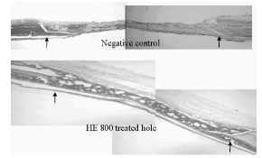
DISTRACTION OSTEOGENESIS STIMULATES INCREASED NUMBERS OF CIRCULATING ENDOTHELIAL PROGENITOR CELLS D. Lewinson1*, S. Bisharat1, A. Rachmiel2 1Dept of Anatomy and Cell Biology, Faculty of Medicine, Technion, Haifa, Israel 2Dept of Oral and Maxillofacial Surgery, Rambam Medical Center, Haifa, Israel Endothelial progenitor cells (EPC) have recently been shown to circulate in increased numbers in several ischaemic experimental animal models and in similar clinical situations as well. The purpose of this study was to look for EPC in a large animal model in which an osteotomoized bone undergoes distraction osteogenesis. We have isolated EPC from the periphral blood of two sheep in which the maxillary bone was osteotomized and the fracture gap was distracted daily for about 2 weeks. The kinetics of the appearance of the EPC in the peripheral blood of the distracted sheep was compared to that of EPC in sham-operated osteotomized, but not distracted sheep. The increase in the percent of EPC from the total white blood cell counts (WC) was 2.5 and 4.5 fold in the two distracted sheep in contrary to no increase in the control sham-operated sheep. Fluorescentically labeled isolated EPC where re-injected to the auotologous animals. They were shown to localize to the callus tissue in ditracted sheep, but not to the callus of control sheep. Their endothelial nature was verified by Tie-2 immunohistochemistry. We suggest to consider ex-vivo expanded autologous EPC as vectors for introducing bone forming genes into bone regeneration sites created by distraction osteogenesis especially in compromized clinical situations. COLLAGEN AS BONE MORPHOGENETIC PROTEIN-1:ITS ROLE IN SKELETAL TISSUES AND ITS FUNCTION UPON ABNORMALITIES M. Tzaphlidou School of Medicine,University of Ioannina, Ioannina, Greece The characteristics of different extracellular matrices derive in large part from the synthesis, assembly, and deposition of collagen molecules and their organization into unique macromolecular structures. These collagen assemblies confer specialized properties to the extracellular matrix in which they are found. The synthesis of collagen molecules and their association to form fibrils requires a number of sequential post-translational events. These include intracellular processes such as hydroxylation and glycosylation, and extracellular ones such as procollagen processing and cross-linking. In bones, it is the procollagen C-proteinase that processes the major fibrillar collagen types I and III, and it may process prolysyl oxidase to the mature enzyme necessary to the formation of covalent cross-links. Type V collagen is a fibrillar collagen of low abundance that is incorporated into and helps regulate the shape and diameter of type I collagen fibrils. As a result a coarse network is formed in the extracellular matrix with thin fibrils. Bone collagen fibrils are much smaller than fibrils from other connective tissues, such as skin, in which collagen is composed by the same types. This difference in fibril diameters may account for the different mechanical properties of the two tissues. Upon skeletal abnormalities, such as osteoporosis, the overall collagen fibril architecture is altered. Such fibrils have a random arrangement in contrast to normal in which fibrils have a characteristic parallel arrangement. In addition, fibril diameter is dramatically affected. In ovariectomized rats, sacrificed 6 weeks after ovariectomy, mean trabecular bone collagen fibril diameter is 47.1 ±3.8 nm while in fibrils from age-matched normal rats is 51.2 ±6.1 nm. The mean values for ovariectomized rats, sacrificed 12 months after ovariectomy, and for the corresponding normals, are 46.1 ±5.5 nm and 53.2 ±4.9 nm respectively. Upon ovariectomy, cortical bone is also suffered of such significant (p<0.001) reduction in collagen fibril diameter. The question of whether skeletal abnormalities are an intrinsic collagen disorder remains to be demonstrated. TWIST INACTIVATION REDUCES CBFA1/RUNX2 EXPRESSION AND DNA BINDING TO THE OSTEOCALCIN PROMOTER IN OSTEOBLASTS M. Yousfi1, F. Lasmoles1, B. Kern2, P. J. Marie1* 1Inserm U349, Paris, France 2Dept Human Molecular Genetics, Baylor College of Medicine, Houston, TX, USA The Saethre-Chotzen (SC) syndrome is characterized by increased osteogenesis and premature fusion of cranial sutures resulting from mutations in TWIST, a basic Helix Loop Helix transcription factor. We previously showed that Twist is an important modulator of human osteoblast proliferation and differentiation. We found that Twist inactivation induced by genetic mutations modulates the expression of osteoblast specific genes such as osteocalcin (OC) and alpha 1(I) collagen (COLIA1). We therefore hypothesized that Cbfa1/Runx2 that controls OC and COLIA1 expression may be a molecular target gene for Twist in human osteoblasts. We tested this hypothesis in Twist mutant (M-Tw) calvarial osteoblasts obtained from a subject bearing the Y103X Twist mutation that introduces a stop codon, leading to a truncated protein without functional bHLH domain. We found that Twist haploinsufficiency in mutant osteoblasts reduced mRNA and protein levels for Cbfa1/Runx2 both during cell growth as well as during in vitro osteogenesis. Moreover, this effect was associated with altered expression of major osteoblast specific genes such as OC, COLIA1, osteopontin (OP) and bone sialoprotein (BSP) whereas osteonectin (ON) was not affected. In addition, electrophoretic mobility shift assay (EMSA) showed reduced-binding ability of Cbfa1 to its target OSE2 element in the OC promoter in mutant osteoblasts. By contrast, Twist inactivation did not hamper Cbfa1 binding on a similar upstream element present in the COLIA1 promoter in mutant osteoblasts. These results indicate that Twist inactivation may control human osteoblast differentiation by altering Cbfa1/Runx2 expression and Cbfa1 binding ability to the osteocalcin promoter, and possibly to OP and BSP promoters. This study provides the first evidence that Cbfa1/Runx2 is a target gene for TWIST in human osteoblasts. A NEW BONE HEALING MATERIAL: A HYALURONIC- CHONDROITIN-HEPARIN-LIKE BACTERIAL EXOPOLYSACCHARIDE Ph. Zanchetta1*, G. Godeau2, K. Senni2, S. Igondjo-Tchen2, N. Lagarde1, J. Guezennec3 1Service Anatomo-pathologie, CHU Morvan, 29200 Brest, France 2Laboratoire de Physiopathologie des Tissus non minéralisés, 1 rue Maurice Arnoux, 92100 Montrouge, France 3IFREMER Dept DRV/VP/BMM. BP 70 29280 Plouzane, France Critical Size Defect (CSD) technique was used to evaluate the bone regeneration capacity of a newly discovered hyaluronic acid like exopolysaccharide synthesized by a bacteria originating from a deep sea hydrothermal vent. A 5 mm diameter hole was made on each parietal bone of male rats. The right hole was filled with either 1 mg of a new bacterial exopolysaccharide referenced HE 800 or with collagen used as negative control, while the left hole remained free of any treatment. After 15 days, the holes and surrounding tissues were examined by direct examination, X-rays, and histological staining. Using HE 800, bone healing was almost complete after only 15 days with osteoblasts lying external bone surfaces and enhancing osteocyte inclusion. Neovascularization was also observed along with an organized trabecular bone. No abnormal bone growth or conjunctival abnormalities were noticed. At the end of the experiment, 95.9 % (SD6.2) bone healing (n=20) was observed (Graphic). Conversely, the collagen treated animals did not demonstrate significant healing 17.8 % (SD18.1). Chemical composition similar to heparin, chondroitin and hyaluronic acid of this polysaccharide could explain the results obtained. Adhesivness and chelation properties may play also an important role.
SYSTEMIC EFFECTS ON BONE HEALING OF AN HYALURONIC ACID-CHONDROITIN-HEPARIN LIKE BACTERIAL EXOPOLYSACCHARIDE Ph. Zanchetta1*, N. Lagarde1, G. Godeau2, K. Senni2, S. Igondjo-Tchen2, J. Guezennec3 1Service d Anatomo-pathologie, CHU Morvan, 29200 Brest, France 2Laboratoire de Physiopathologie des Tissus non minéralisés, 1 rue Maurice Arnoux, 92100, France 3IFREMER Dept DRV/VP/BMM. BP 70 29280 Plouzané, France Critical Size Defect (CSD) technique was used to evaluate the systemic activities on bone regeneration capacity of a newly discovered hyaluronic acid-chondroitin- heparin like exopolysaccharide synthesized by a bacteria originating from a deep sea hydrothermal vent. Some systemic effects were previously detected on earlier experiments. A 5 mm diameter hole was made on each parietal bone of male rats. The right hole was filled with 0.5 mg of a new bacterial exopolysaccharide referenced HE 800, while the left hole remained free of any treatment. After 21 days, the holes and surrounding tissues were examined by direct examination, X-rays, and histological staining. Using HE 800, bone healing was almost complete after only 21 days in the treated hole and always complete in the control side by a supposed systemic effect. Neovascularization was also observed along with an organized trabecular bone on both sides. No abnormal bone growth or conjunctival abnormalities were noticed. At the end of the experiment, 90.1 % (SD 5.2) bone healing (n=20) was observed on the treated side; conversely, the control side animals demonstrate an amazing healing 100% SD 0.5) by a systemic effect.
WITHDRAWN EXPRESSION OF SEMAPHORIN-3A AND ITS RECEPTORS IN BONE: POSSIBLE ROLE IN SKELETAL DEVELOPMENT C. Gomez*, B. Burt-Pichat, L. Malaval, C. Chenu INSERM Unit 403, Hôpital E. Herriot, Pavillon F, 69437 Lyon Cedex 03, France Recent studies have shown evidence for a neural regulation of bone development and remodeling. However, the molecules and mechanisms involved in the development and dynamic maintenance of bone innervation are unknown. During the development of the central nervous system, extending axons are oriented to their targets through the actions of different families of guidance molecules. We investigated the expression of Semaphorin 3A (Sema 3A) and its receptors in in vitro models of bone cell differentiation and in developing bone. RT-PCR analysis was performed at successive stages of osteoblast differentiation in newborn rat calvaria cell cultures. These experiments, completed by immunolabelling, showed the expression by osteoblastic cells of Sema 3A, as well as its receptors, Neuropilin-1 (NP-1) and -2, Plexins A1 and A2. Sema 3A and NP-1 were expressed throughout the osteogenic differentiation sequence, with little regulation except for an increase of NP-1 mRNA in differentiated stages. Only Plexins and Neuropilins were present in osteoclasts differentiated from RAW 264.7 cells. We studied the expression of Sema 3A and NP-1 in vivo using immunocytochemistry on rat long bone sections at gestational days (GD) 17 to 21, and 2 and 3 weeks post natal. At early stages of endochondral ossification (GD 17-19), both proteins were present in the periosteum and in chondrocytes of the mid-diaphysis before (GD17) and after vascular penetration (starting on GD18) and early primary ossification. The same labelling was observed in epiphyseal chondrocytes before and after the onset of secondary ossification. At day 21, NP-1 was still expressed in the periosteum, contrarily to Sema 3A. Both molecules were later detected in pre-hypertrophic and hypertrophic chondrocytes of the growth plate and in osteoblasts lining bone trabeculae. RT-PCR analysis was performed on differentiating cultures of the chondrocytic cell line MC615, confirming the expression of Sema 3A and NP-1 by this cell type. Our results demonstrate the expression of Sema 3A and its receptors in bone cells in vitro and in vivo during bone growth. Their distribution pattern, temporally and spatially preceding the onset of bone tissue formation, suggests that Sema 3A signalling may play a role in the regulation of skeletal development. EFFECTS OF TCDD ON METAPHYSEAL BONE IN A TCDD- SENSITIVE AND A TCDD-RESISTANT RAT STRAIN N. Stern1*, M. Lind1, M. Viluksela2, J. T. Tuomisto2, J. Tuomisto2, H. Hĺkansson1 1Institute of Environmental Medicine, Karolinska Institutet, Stockholm, Sweden 2Department of Environmental Health, National Public Health Institute, Kuopio, Finland Introduction Bone development and maintenance are strictly controlled by hormonal interactions. Dioxins are known to disturb several hormonal systems, inter alia to have antiestrogenic effects. Therefore dioxin exposure may cause trabecular bone loss and decrease of bone strength at sites containing both trabecular and cortical bone such as long bone metaphysis. Sensitivity to dioxin-induced toxicity is highly variable. An animal model based on different sensitivity to various endpoints of dioxin toxicity has previously been developed. Long-Evans (L-E) is the most TCDD-sensitive rat strain, Han/Wistar (H/W) is the most resistant. In a recent study L-E rats demonstrated higher sensitivity than H/W rats to TCDD-induced changes in diaphyseal geometry and strength of tibia. In this study we used L-E and H/W rats to examine the effects of TCDD on cortical and trabecular bone of femur metaphysis. Material and Methods Female L-E and H/W rats were given a total dose of 0, 0.17, 1.7 and 170 (H/W only) mg/kg TCDD s.c. weekly during 20 weeks. The right femur was dissected and the length was measured. The femoral metaphysis was scanned at a point distanced 20% of the bone length from the distal end of femur with a pQCT system (Stratec XCT 960A, Birkenfeld, Germany) with a voxel size of 0.148 mm3. A 0.400 cm-1 attenuation threshold was set as a lower limit to define the trabecular bone region. The trabecular bone mineral density and area, as well as cortical/subcortical bone density and area were analysed. The Mann-Whitney Rank Sum test was used with a significance level of p<0.05. Results and Discussion Cortical/subcortical area of distal femur metaphysis was smaller in L-E rats at 1.7 and 17 microg/kg and in H/W rats at 17 and 170 microg/kg dose levels (Fig 1). Trabecular bone mineral density was decreased in LE rats at 17 microg/kg vs. corresponding controls (Fig 2), but not in H/W rats. The data show that TCDD exposure causes trabecular bone loss and confirms previous findings that dioxins interfere with bone remodelling.
BIOPHYSIOLOGY OF AUTOGENIC BONE GRAFT. ALP, QCT AND HISTOLOGIC SHORT-TERM STUDY A. Smailagic1*, A. Redji 2, B. Hadjihasanovic3, S. Lappalainen4 1University Clinic Center Sarajevo Clinic for Maxillofacial Surgery, Bosnia and Herzegovina 2Faculty of Medicine, Institute for Human Genetic and Biology, University of Sarajevo, Bosnia and Herzegovina 3University Clinic Center Sarajevo Radiologic and Diagnostic Center, Bosnia and Herzegovina 4 BonAlyse Oy, Jyväskylä, Finland Autogenic bone grafts are currently the most widely used method in skeletal surgery for reconstructive purpose. Certain problem with extensive resorbtion (up 50%), sequestration, inadequate integration with host bone can compromise success. Factors as rigid fixation, embriologic origin of transplant, blood supply and presence of growth factors are some of reason transplantation procedure success. White New Zealand rabbit underwent reconstruction of unilateral created non critical size defect of mandible, fixed with mini plate and bridged with autogenic bone graft from spine scapule. Result were evaluated measurement of ALP activity, QCT GEANIE 2.1 (BonAlyse Oy, Jyväskylä, Finland) program of bone analysing and clinical and histologic examinations.Result: ALP activity was significancy higher after 30 days, density of transplant after 30 days was 555-696 HU (GEANIE 2.1 analysing), and clinical inspection and histologic analysing showed bridged defect and complete integation of graft with host bone and new osteoblast, osteocit and angiogenesis. Conclusions: Significant higher ALP activity after 30 days indicate on time beginning of process osteoinduction and osteoblast differentiation. Histologic and clinic inspection indicate that after 45 days bone graft are total revasculated and integrated with host bone. Autogenic graft showed no immune response and viral transmission. Different embriogenic origin (intramembranose) of recipient bone-mandible and enhondral origin of graft from spina scapule showed no implicacations on osteointegated process in short term study. OVERLOADING OF THE TEMPOROMANDIBULAR JOINT UPREGULATES THE JNK/AP-1 SIGNALING PATHWAY TRIGGERING CHONDROBLASTIC DIFFERENTIATION D. J. Papachristou1,2*, P. Perttiniemi3, T. Kantomaa3, N. Agnantis2, A. G. Papavassiliou1, E. K. Basdra4 1Department of Biochemistry, University of Patras School of Medicine, Patras, Greece 2Department of Pathology, University of Ioannina School of Medicine, Ioannina, Greece 3Institute of Dentistry, University of Oulu, Oulu, Finland 4Department of Orthodontics, University of Heidelberg, Heidelberg, Germany Mechanical loading has been long recognized as an important regulatory factor in bone and cartilage homoestasis and a determinant of skeletal morphology. However, the molecular mechanisms that govern the response of chondroblasts to mechanical stimulation remain elusive. The transcription factor c-Jun together with members of the Fos family proteins (c- Fos, FosB, Fra1/2) are major components of the AP-1 (activator protein-1) transcription complex. Recent in vitro studies have indicated that AP-1 plays an important role in chondrogenic gene regulation as well as chondroblastic differentiation and proliferation. The aim of the present study was to explore the effect of increased temporomandibular joint loading on the proliferation and differentiation of condylar cartilage chondrocytes, in vivo. To this end, 100 rats were assigned to two groups: the first group was fed soft diet (which simulates normal masticatory movements), while the second group was fed hard diet that causes increased temporomandibular joint loading. Biopsy sections from temporomandibular joint of both groups were obtained at 2, 6, 12, 24, and 48 hours after the experiment initiation. We employed immunohistochemical staining analysis to investigate the in situ expression of c-Fos, in correlation to cellular levels of pc-Jun, the phosphorylated (hence activated) form of c-Jun. Moreover, the expression and activation profile of JNK2 (c-Jun N-terminal kinase 2), the principal kinase targeting c-Jun, was examined. The articular cartilage of the first group displayed nuclear immunoreactivity for c- Fos, that was gradually increased as the experimental procedure evolved. The expression levels of c-Fos were accompanied by a co-localized enhancement of pc- Jun. Augmented levels of JNK2 and its phosphorylated/activated form, p-JNK, were observed at 24 and 48 hours after experiment initiation. In the second group, immunoexpression of the aforementioned proteins was also increased as the experiment progressed. However, expression levels of the examined proteins were significantly higher in the second, compared to the first group, at 12, 24, and 48 hours after the experiment initiation. Our results suggest that mechanical loading potentiates the JNK/AP-1 signaling pathway and pose a novel mechanism that might be implicated in the chondroblastic differentiation process. THE EXTRACELLULAR MATRIX 1(ECM1) GENE IS ESSENTIAL FOR MOUSE EMBRYOGENESIS J. Liekens1*, J. Merregaert1, P. Smits1, L. Umans2, D. Huylebroeck2, A. Zwijsen2 1Lab of Molecular Biotechnology, Dept. of Biomedical Sciences, University of Antwerp, Universiteitsplein 1, B-2610 Wilrijk, Belgium 2Department of Cell Growth, Differentiation and Development, (VIB), University of Leuven, Campus Gasthuisberg, Herestraat 49, B-3000 Leuven, Belgium The Extracellular matrix gene 1 encodes for a glycoprotein of 559 amino acid residues with a typical cystein CC-(X7-10 )-C distribution. This cystein arrangement forms 'double loop' domains similar to those of the serum albumin protein family (1). During embryogenesis Ecm1 mRNA was detected in close association with specific sets of developing blood vessels at different stages, particularly during midgestation. Its distribution there is very similar to that of flk-1. However,unlike flk-1, Ecm1 mRNA expression was downregulated before birth. The biological function of Ecm1 encoded protein(s) is still unknown. A role for Ecm1 in processes such as epidermal differentiation (2), angiogenesis (3) and endochondral bone development (4) has been suggested. Recently, ECM1 was linked to the rare human autosomal recessive disorder Lipoid proteinosis (5). To understand the function of Ecm1, we used homologous recombination in mouse embryonic stem cells to produce Ecm1 null mice by deleting the first 2 exons of the mouse Ecm1 gene thus deleting the start of transcription and of translation. Mice heterozygous for the Ecm1 null mutation (Ecm1 +/-) are fertile and grossly indistinguishable from wild type. Mice homozygous for the Ecm1 null mutation (Ecm1 -/-) are not viable and the mutants die around gastrulation. Analysis of the embryo's of heterozygous matings revealed that the embryonic lethality occurs between 4.5 dpc and 6.5 dpc. Further characterization is currently being performed. The phenotype demonstrates a crucial role for Ecm1 in the early stages of embryogenesis that cannot be compensated for at that time. In order to assess the putative role of Ecm1 during endochondral bone formation transgenic mice overexpressing Ecm1 in cartilage by the chondrocyte-specific cis- element of the alfa1(II) procollagen gene have been generated. The impact of Ecm1 on terminal chondrocyte differentiation and growth plate tissues is currently under investigation in these animals. References: 1) Bhalerao et al. (1995) J. Biol. Chem. 27, 16385-16394 2) Smits et al., (2000) J Invest Dermatol 114:718-724, 3) Han et al, (2001) FASEB J. 15,988-994, 4) Deckers et al., (2001), Bone ,28, 14-20 5) Hamada et al., (2002) Hum. Mol. Genet. 11, 833- 840. APPETITE-DEPENDENT EFFECT OF HYPOXIC HYPOXEMIA ON BIOMECHANICAL PROPERTIES OF THE GROWING RAT FEMUR R. M. Alippi*, M. I. Olivera, C. Bozzini, C. E. Bozzini Department of Physiology, Faculty of Odontology, University of Buenos Aires, Argentina We have previously shown in growing rats that hypoxemia induced by exposure to hypobaric air exerts a negative effect on body and longitudinal skeletal growth, which is accompanied by alterations in the biomechanical performance of bones. These manifest in diminished stiffness and lower than normal resistance to fracture. We have also shown that hypobaric hipoxia inhibits appetite and, as a consequence, food intake. The aim of the present study was to separate the above-cited effects of hipoxia per se from those induced by nutritional impairment by performing pair-feeding studies. Three groups of 7 female Wistar rats aged 26 d were established as follows: CNx rats were maintained in room air and taken as normoxic controls. Hx rats were continuously exposed to air maintained at 506 mb in a simulated high altitude chamber for 60 d. PFNx rats were treated as CNx ones but food was offered to them in the amount freely eaten by Hx animals. Body weight and length, as well as food consumption, were recorded daily. At the end of the study period, rats were sacrificed by ether overdose. Morphometric analyses were done on the left femur. The right bone was biomechanically tested by the three-point bending test and its bone mass assessed by dual energy X-ray absorptiometry (DEXA). Both femoral weight and length were lower in both Hx and PFNx rats than in Nx ones. The following effects were observed in the right bone of the former groups: 1) diminished bone mass (BMC) and bone mineral density (BMD); 2) diminished resistance to fracture (Wf); 3) reduction in bone stiffness (Wy/dy); and 4) impairment of bone material quality (Young's modulus, E). The negative effects of treatments on BMC and BMD were higher in Hx than in PFNx rats. It can be concluded that hypobaric hypoxemia negatively affects the quantity as well as the quality of femoral bone. The effects are not entirely attributable to the lowered food intake that usually accompanies the hypoxic states. ASSESSMENT OF BONE MINERAL DENSITY OF THE CALCANEUS IN HEALTHY SWEDISH 7-YEAR OLD CHILDREN BY DXL CALSCAN A-C. Söderpalm1,3*, R. Kullenberg2, K. Albertsson Wikland3, D. Swolin-Eide3 1Dept. of Orthopaedics SU/Östra Hospital, Göteborg, Sweden 2Dept. of Radiology, Halmstad Hospital, Halmstad, Sweden 3Göteborg-Pediatric Growth Research Center,Queen Silvia Children's Hospital, Sahlgrenska Academy at Göteborg University, Göteborg, Sweden A debated issue in bone research is the optimal techniques to determine the bone mass in growing children. Dual X-ray and Laser (DXL) Calscan measures areal bone mineral density (BMD) by using dual X-ray absorptiometry in combination with laser measurement of the total heel thickness. This technology reduces the uncertainty related to variable composition of soft tissue in adults. The DXL Calscan is portable, easy to use, has a short measurement time and gives a low absorbed dose (<0.12microSv). Aim: To investigate: 1) if the device was tolerated by children 2) if BMD could be measured with good accuracy 3) if BMD was related to height, weight and body mass index (BMI) at the time of measurement and 4) to create a reference material in 7y old children in a cross-sectional study. Methods: The DXL Calscan was modified for children with a lower absorbed dose and adapted software. 112 healthy children (57 boys, 55 girls, mean age 7.5y) were included. The left foot was scanned, the actual length, weight and foot length was measured. A questionnaire comprised physical activity, milk intake, osteoporosis in relatives, weight and height at 1y of age. Results: The intra-individual CV measured by 2 repeated measurements on 27 subjects was 2.44% for BMD and 2.61 % for bone mineral content (BMC). The mean values for the weight of the subjects was 27.1±5.4 kg, length 127.2±5.7 cm, BMI 16.6±2.3 kg/m2, foot length 197.9±11.5 mm, calcaneus thickness 40.4±3.4 mm and calcaneus height 33.8±2.5 mm. The mean BMD in the subjects was 0.30±0.05 g/cm2 and BMC 0.22±0.04g. Girls showed higher BMD than boys (p<0.05). BMD was significantly correlated to weight, BMI, foot length, height and calcaneus height (p=0.001). Weight at 1y correlated to BMD (p= 0.016). No significant correlation was found between BMD and physical activity, milk intake and osteoporosis in relatives. In conclusion, the measurements were well tolerated and easily performed and this study shows reference values for BMD in calcaneus in 7y old Swedish children. Further studies are required to evaluate this method. LEUKEMIA INHIBITORY FACTOR (LIF) IS EXPRESSED IN HYPERTROPHIC CHONDROCYTES AND VASCULAR SPROUTS DURING OSTEOGENESIS E. Grimaud1, F. Blanchard1, C. Charrier1, F. Gouin2, F. Rédini1, D. Heymann1* 1Labo Physiopathologie de la Résorption Osseuse, Faculté de Médecine, Nantes, France 2Orthopaedic Department, Nantes Hospital, Nantes, France Endochondral ossification is closely controlled, especially by cytokines and growth factors. Leukemia inhibitory factor (LIF), a member of the gp 130 cytokine family, is involved in osteoarticular tissue metabolism, is expressed by hypertrophic chondrocytes from cartilage-forming tumors of bone. It also modulated proteinase production and is a chemoattractant for endothelial cells. To investigate its role during endochondral ossification, LIF expression was then analyzed immunohistochemically in articular tissues of rats and humans. Sprague Dawley rats were used to study LIF expression in growth cartilage during bone formation. Age groups (6 animals each) of newborn (day 0 to 21) and 7-, 9- and 12-week-old adult rats were investigated. Five femurs with osteogenesis imperfecta were obtained from human fetuses after therapeutic abortion at 15, 21, 23, 24 and 25 weeks of amenorrhea. Normal human articular cartilages harvested on epiphysis resected because of metaphyseal bone tumors were used as a control. At day 0, LIF immunostaining was observed in hypertrophic chondrocytes, with the strongest signal in the lower hypertrophic zone. LIF localization was essentially cytoplasmic. The other cartilaginous zones of the growth plate showed no positive staining. Staining was also observed in osteoid zone in the bone formation area. Similar results for LIF localization were obtained in newborn rat femurs at day 1 to 6. At day 14, vascular sprouts invaded cartilage in the middle of the epiphysis, forming ossification centres. Immunostaining showed that LIF was localized in the cytoplasm of hypertrophic chondrocytes from diaphyseal and epiphyseal ossification centers. Other zones of bone formation (resting, proliferative, maturation) and calcified matrix were negative. Positive LIF staining was also detected in mesenchymal cells of the vascular sprout and in osteoblasts located in zone of bone remodelling whereas osteoclasts were negative. Positive LIF staining was no longer observed in adult rat femur (week 7, 9, 12). LIF was not observed in adult articular cartilage and bone marrow. Similar results were obtained from rat and human samples. These results and other data in the literature suggest that LIF is involved in the delicate balance between the rate of formation of calcified cartilage and its vascularization for bone development. DENTINAL TISSUE:ULTRAMICROSCOPIC OBSERVATION M. Baldi*, M. Frascaria, M. Ponzi, D. Battisti, M. Giannoni School of dentistry, University of L'Aquila, Italy Conditioning and treatment of dental tissues is the objectives in lots of researches, to find the best standard procedures.We usually use SEM to observe histologic aspects of these tissues, but the metalisation of specimen, that this instrument needs, doesn't allow to analyse tissues before and after conditioning. In our study, we use a different kind of SEM with controlled pressure, that allows, in some conditions, to avoid metalisation. Conditioning agents were Orthophosphoric acid 37%, used as conditioner of enamel and dentin in preparation of composite fillings (Etching process), EDTA 17%, utilised as smear layer remover, and sodium ipochloride, used in endodontic therapies. Etching process with orthophosphoric acid allows bond between adhesive and enamel, making possible the penetration of bonding adhesive in dentine's tubuli and realize of bonding system of composite fillings. Orthophosphoric acid as been used for 15 sec, EDTA for 60 sec. and ipochloride for 3 min at the concentration of 5%.Fifteen bicuspids, catched for orthodontic and periodontal problems, were sectioned and prepared to obtain fractured and abraded surfaces, and each kind has been treated with the tree conditioning agents described above. We observed specimens before and after the treatments with a SEM Philips XL 30CP. The images obtained by SEM with pressure control, showed the action of orthophosphoric acid and EDTA on smear layer. There were big differences between fractured surfaces and abraded ones, especially in smear layer thickness. Sodium ipochloride showed no action on smear layer, as expected. Our results has been completely similar to ones still obtained from other investigations. Although the imaging quality of this kind of SEM is not comparable with ones obtained with traditional SEM, considerable the possibility to observe the same sample after a peculiar treatment, that is not possible in other ways. We believe that methods we used in this study could be considered in the future, together with traditional SEM, to reach more informations about these treatments. EFFECTS OF THE ANTI-INFLAMMATORY CYTOKINES IL-4 AND IL-13 IN THE DOUBLE KO MICE A. Frost1*, C. J. Silfverswärd1, C. Ohlsson2, O. Nilsson1 1Inst Surgical Sciences, Uppsala Universitet, Uppsala, Sweden 2Dept of Internal Medicin/Endocrinology, Sahlgrenska University Hospital, Gothenburg, Sweden Inflammation in, or in the vicinity of bone can cause osteolysis or osteosclerosis through the actions of secreted inflammatory mediators. We have previously shown that the anti-inflammatory cytokines IL-4 and IL-13 inhibit proliferation of and stimulates IL-6 secretion by human osteoblast in vitro. To study the physiological role of these cytokines we used the IL-13 (-/-) knockout mouse and the IL-4/-13 (--/--) double knockout mouse. The skeletal phenotypes of these transgenic mice were studied with DEXA and QCT and biomechanical testing was performed by tree point bending. The mice were analysed in adolescence at 6 weeks of age and when sacrificed at 12 weeks of age. There was no significant difference in cortical or trabecular bone parameters in 6 week-old IL-13 (-/-) or IL-4/-13 (--/--) mice compared to control mice or when comparing the two knock-out lines. In 12 week-old mice we found a significant thinner cortical bone and a more fragile bone in the male, but not in female IL-4/-13 (- -/--) mice compared to the controls. The effect of the depletion of the anti-inflammatory cytokines IL-4 and IL-13 on cortical bone seems to dependant on the inability of Th 2 cells to synthesise IL-4. IMPLANTATION OF OCTACALCIUM PHOSPHATE (OCP) COMBINED WITH BONE MATRIX GELATIN (BMG) INDUCED BONE REGENERATION IN RAT SKULL DEFECTS F. Sargolzaei1*, A. Sobhani2, M. Akbari2, B. Niknafs3, A. Hedayat Poor2 1Dept. of Anatomy, Faculty of Med., Zahedan Univ. of Med. Sci., Iran 2Dept. of Anatomy, Faculty of Med.Tehran Univ. of Med. Sci., Iran 3Dept. of Anatomy, Faculty of Med., Tabriz Univ. of Med. Sci., Iran Abstract: Introduction:In order to evaluate bone induction and repair in cranial bone defects by the use of combination of OCP/BMG. Methods and Materials: we used 40 young male Sprague dawley rats (5-6 weeks age). A full thickness standardized trephine defect, 5mm in diameter, was made in the rat parietal bone and OCP combined with BMG was implanted into the defect. No OCP/BMG particles were implanted in control group. On the 5th, 7th, 14th, and 21st days after implantation, the specimens were harvested. After processing the tissues by routine histological procedures, we prepared 5 micrometer sections of the samples, stained with H& E and alcian blue. Finally the sections were studied by light microscope. Results: On the 5th day after implantation, we observed inflammatory cells around the implanted material, especially around the OCP particles. A few clusters of cartilage cells were observed on the 7th day, which located between the BMG particles in the central position of defect. On the 14th day after implantation osteogenesis was initiated from the margin of the defect. In addition to bone formation from the margins toward the center, we observed more appositions of new bones around the implanted materials. At the end of 21st day, almost all of the OCP/BMG particles were absorbed and bone trabeculae, bone marrow cavities and bone marrow tissues were appeard. In control group, at the end of 21th day, a few cluster of new bone were observed near to the defects margin and host bone. Conclussion:As the findings show, combination of OCP/BMG could stimulate bone induction and new bone formation in bone defects, so it seems that these biomaterials could be used in the repair of cranial bone defects. Key word: Bone regeneration, Octacalcium phosphate, Bone matrix gelatin, Parietal bone, Rat. THE REPAIR OF CRANIAL BONE DEFFECT BY USING OF ENDOCHONDRAL BONE MATRIX GELATIN (EC BMG) IN RAT A. Sobhani1*, F. Sargolzaie2, H. Rafighdoost2, M. Akbari1, I. R. Kashani1, M. Abbasi1 1Dept. of Anatomy, Faculty of Med., Tehran Univ. of Med. Sci. Tehran, Iran 2Dept. of Anatomy, Faculty of Med., Zahedan Univ. of Med. Sci., Tehran, Iran Abstract: Introduction: Many investigators have used bone matrix gelatin (BMG) for bone induction intramuscularly and subcutaneously. A literature review revealed only a few studies about the process and type of ossification by BMG in skull bone defects. Aims of investigation: Evaluation of Ec BMG effects on repair of cranial bone defects. Materials and Methods: Ec BMG was prepared as previously described by Urist. The defects were produced with 5-mm diameter in parietal bones and filled by BMG particles. No BMG was used in control groups. For evaluation of new bone formation and repair, the specimens were harvested on days 7, 14, 21 and 28 after operation. The samples were processed histologically, stained by H&E, Alizarin red s staining and Alcian blue and studied by a light microscopy. Results: In control groups: Twenty-eight days after operation a narrow rim of new bone was detectable attached to the edge of defect. In BMG groups: At day 7th after operation young chondroblast cells appeared in whole area of defect. At day 14th after operation hypertrophic chondrocytes showed by Alcian blue staining and calcified cartilage were detectable by Alizarin red s staining. The numerous trabeculae spicules, early adult osteocyte and highly proliferated red bone marrow well developed on day 21. Finally, typical bone trabeculae with regulated osteoblast cells and some osteoclast cells were detectable at day 28 after operation. Discussion: Ec BMG could stimulate bone induction in cranial bone defects by the way of endochondral ossification. Although, some of researchers haven't believed this idea. Conclusion: According to results of this research, Ec BMG could be a good biomaterial substance for new bone induction in bone defects. Key Words: Bone Induction, Bone Matrix Gelatin (BMG) and Rat. APO-1 AND CASPASE-8 ARE DISTINCT EXPRESSED IN THE HUMAN GROWTH PLATE K. Trieb*, E. Cetin Dept. of Orthopedics, University of Vienna, Austria Enchondral ossification, the replacement of cartilage by bone in the human growth plate (physis), is the basis of longitudinal bone growth. Former animal studies showed that chondrocytes in the growth plate are assumed to undergo apoptosis, a form of programmed cell death. The aim of our study was to investigate the role of APO-1 and Caspase-8, two apoptosis-mediating proteins, in chondrocytic maturation and differentation in the physis by the technique of immunohistochemistry (IHC), which, to our knowledge, was carried out the first time. We used the human growth plates of resected polydactylic fingers of five infants (f:m=1:4; mean age: 16,6 months; range: 13-22 months). Control sections were incubated with Anti-D antibodies. In each zone (RZ, PZ, HZ) of one section, positively and negatively stained cells were counted. Statistical analysis was done by Kruskal-Wallis test. APO-1 positive and Caspase-8- positive chondrocytes were found in all three investigated zones (RZ, PZ, HZ) of the physis. The expression of the proteins increased from the RZ to the HZ near to closure of the growth plate. 13,17 (SD1,8)% cells in the RZ were positive for Caspase-8, 19,33 (SD 2,91)% in the PZ and 28,22 (SD 2,13)% in the HZ (p<0,001). APO-1 was expressed in 17,4 (SD 6,17)% in the RZ, in 22,1 (SD 6,2)% in the PZ and in 33,07 (SD 8,39)% in the HZ (p<0,001). The findings of this study indicate that both APO-1 and Caspase-8 have a distinct expression pattern during chondrocyte differentation and further a significantly increasing expression in the HZ. Our data suggest that proapoptotic proteins are involved in the growth plate regulation of human chondrocyte maturation. BONE STRENGHT IN NEWBORNS L. Pedrotti1*, G. Tuvo1, B. Bertani1, R. Mora1, L. Rosti2, A. Mastretti3, S. Quaglini4 1Dept of Orthopaedic and Traumatology, University of Pavia, Pavia, Italy 2Dept of Neonatology, Istituto di Cura Citta' di Pavia, Pavia, Italy 3Medical Director, Istituto di Cura Citta' di Pavia, Pavia, Italy 4Dept of Computer Science and Systems, University of Pavia, Pavia, Italy It is crucial to monitor the bone healthness during the growth. The bone takes its shape through cycles of resorption and apposition of new bone. Alterations in these processes may cause bone loss as well as structural instability thus affecting the peak of bone mass (PBM). It is also well known that the bone strenght is tightly related to the mineral content as well as to elasticity, microarchitectural structure and cortical thickness. Bone strenght evaluation using DXA is not reliable in children because this technique is based upon measurement of the bone area which depends on physical and skeletal size, and above all DXA reflects the mineral content only. The ultrasound devices are easy to be used, unexpensive and, primarly, they do not require the use of Xrays. For all these reasons ultrasound based devices are used in order to analyse the bone health in paediatric population. In this study a cluster of 220 newborns underwent bone strenght evaluation using the OmnisenseTM device (Sunlight Technologies). The measurements have been done at the medium third of the tibial shaft using a specific probe that is reliable in premature babies too. The accuracy test have been performed in 20 newborns measuring the bone mass in the 1st, 2nd, and 3rd day after birth. The statistical analysis has been done comparing the bone mass with the following parameters: weight, length, thoracic and cranial circumference, duration of the pregnancy, type of delivery and Apgar index. COMPARISON OF THE EFFECTS OF ENDOCHONDRAL BONE MATRIX GELATIN (EC BMG) WITH AUTOGENOUS BONE GRAFTS IN THE RECONSTRUCTION OF BONE DEFECTS: AN EXPERIMENTAL STUDY IN RABBITS H. Shahoon1*, M. Bayat2, A. Sobhani3, B. Eslami4 1Dept. of Maxillofacial surgery, Faculty of Dentistry, Shahid Behshti Univ. of Med. Sci., Iran 2Dept. of Maxillofacial surgery, Faculty of Dentistry, Tehran Univ. of Med. Sci., Iran 3Dept. of anatomy, Faculty of Medicine, Tehran Univ. of Med. Sci., Iran 4Dept. of Oral Pathology, Faculty of Dentistry, Shahid Behshti Univ. of Med. Sci., Iran Abstract: Purpose: The use of autogenous bone grafts is usual for the reconstruction of bone defects in routine practice. The problem of producing and use of grafts have encouraged investigators to search for a suitable alternative, of which, Bone Matrix Gelatin (BMG) is the most popular material. In order to evaluate and compare the effect of BMG and autograft on the reconstruction of bone defects, this study was conducted in rabbits. Materials and Methods: The design of the study was experimental and data was collected using a questionnaire and microscopic observation. Male white New Zealand rabbits were used for this study. Using Urist technique, BMG was prepared and grafts were also removed from rabbits left iliac bone. The grafts were placed on maxilla right defect and BMG was applied to fill left defect. The rate and amount of reconstruction were compared at 7th. 14th, 24th and 60th days after operation according to following items: 1- Type of osteogenesis 2- Cartilage formation 3- Rate of bone formation 4- Inflammation types. Results: The results showed that through time the inflammation degree was reduced and bone formation was increase in two groups. Conclusion: This indicates that Ec BMG, like autograft, can be useful in the reconstruction of rabbit's masillofacial bones defects. Key words: Endochondral Bone Matrix Gelatin (Ec BMG), Autograft, Bone Defects and Rabbit. ULTRASTRUCTURAL RESEARCH OF THE BONE MINERAL AT FILLING BONE DEFECTS BY DALARGIN V. I. Luzin*, K. P. Garboos, M. V. Nechoroshev, M. G. Grishchuk State Medical University, Lugansk, Ukraine Experiment is carried out on 72 white rats with initial weight of 130-150 grams. Of 24 animals under aether narcosis a standard pine forest in diameter of 2,2 millimeters rendered foraminous through defect on border proximal metaphysis and diaphysis both tibial bones which filled by dalargin (experimental group). 24 animals filling bone did not make (1 control group). As 2 control group served 24 intact rats. Later 15, 30, 60 and 90 days after operation of animals were took out from experiment under an aether narcosis. For the further research allocated fragments of tibial bones, the places with defect were tooking, and pounded them in a powder and investigated by X-ray diffraction method. Have established, that at introduction dalargin in a zone of defect the sizes of elementary cells a new formated bone mineral along an axis C were by 15 and 90 days of experiment were authentically less than control values accordingly on 0,13% and on 0,18%. It testifies to active formation and growth of hydroxylapatite elementary cells. The parameters of elementary cells are the type of mineral organization and this fect characterized by the small changes in the amplitude of deviations. The sizes of blocks of coherent dispersion (that is conglomerates of elementary cells) as were less control: on 10,58% by 15 day, on 6,22% by 30 day and on 5,18% by 90 day. This fact testifies to increase in the common exchange surface of a bone mineral. At last, the microtexturation factor (uniformity of orientation of crystals in a lattice), designed on a method of a ratio of reflexes changed as follows: by 15 day it was less than control value on 20,03% that meets to active formation of roughly fibrous bone fabric. Then the factor microtexturation increased and by 90 day was more than control values on 28,81%. This fact testifies to a high degree of orderliness of again generated crystal lattice. Thus, at introduction in a zone of bone defect of a dalargin, the ultrastructure of a formed bone mineral is optimized. It is shown in increase in the common exchange surface and ordering crystals lattices bone hydroxylapatite. POOR MAINTENANCE OF HIGH BONE DENSITY AFTER CESSATION OF ICE HOCKEY CAREER: A SIX YEAR LONGITUDINAL STUDY IN MALES A. Gustavsson1,2, T. Olsson1, P. Nordström2,3* 1Department of Public Health and Clinical Medicine, Umeĺ University, Umeĺ, Sweden 2Sports Medicine Unit, Department of Surgical and Perioperative Sciences, Umeĺ University, Umeĺ, Sweden 3Department of Geriatric Medicine, Umeĺ University, Umeĺ, Sweden To optimize peak bone mass by intense physical activity during childhood and adolescence could decrease the risk of osteoporosis, given that the bone gain is not lost when the activity is decreased later in life. In this longitudinal study 43 ice hockey players (aged 16.7±0.6) and 25 control subjects (aged 16.8±0.3), were measured at baseline and after three and six years. The groups were not significantly different in age, weight, or height. Bone mineral density (BMD, g/cm2) was measured in the total body, femoral neck, and spine using dual-energy X-ray absorptiometry (Lunar DPX- L). Volumetric bone density (vBMD mg/cm3) of the femoral neck was estimated. The ice hockey players were found to gain significantly more femoral neck BMD (0.07 vs. 0.03 g/cm2, p=0.04) and femoral neck vBMD (16 vs. 0 mg/cm3, p=0.049) than the controls between the first and second visit. At the second visit the ice hockey players were found to have significantly higher BMD at the femoral neck and total body compared to the controls (p<0.05). Between the second and third visit, 21 ice hockey players stopped their active career. These men lost significantly more femoral neck BMD (0.10 vs. 0.02 g/cm2, p<0.001) and femoral neck vBMD (38 vs. 4 mg/cm3, p<0.001) compared to the ice 22 hockey players who continued the training. The former ice hockey players also lost significantly more neck vBMD (38 vs. 14 mg/cm3, p=0.009) compared to the controls during the same period. At the third visit, at 23 years of age, only the 22 ice hockey players who had continued their training were found to have significantly higher BMD at femoral neck (p=0.01), total body (p=0.04), and spine (p=0.02), compared to the controls. During this study the changes in training were highly associated with the changes in BMD at all sites in the 43 ice hockey players (r=0.44-0.46, p<0.01). In summary, we have demonstrated a fast BMD loss at the femoral neck with decreased training in young men. These data suggest that a physically active lifestyle during childhood and adolescence may not prevent osteoporosis of the femoral neck later in life. THE EFFECT OF OCTACALCIUM PHOSPHATE (OCP) ON THE REPAIR OF PARIETAL BONE DEFFECT IN RAT B. Niknafs1*, F. Sargolzie2, A. Sobhani3, M. Akbari3 1Dept. of Anatomy, Faculty of Med., Tabriz Univ. of Med. Sci., Iran 2Dept. of Anatomy, Faculty of Med., Zahedan Univ. of Med. Sci., Iran 3Dept. of Anatomy, Faculty of Med., Tehran Univ. of Med. Sci., Iran Abstract: Introduction: Hydroxyapatyte and its derivatives used extensively by many researchers as osteoconductor factors. But, some of the researchers were believed that octacalcium phosphate is an osteoconductor. Aims of investigation: Evaluation of octacalcium phosphate effect on repair of cranial bone defects. Materials and Methods: The OCP was prepared by Legeraus method. A defect with 5-mm diameter was produced and 5mg OCP was implanted in parietal bones. On days 7th, 14th and 21st after operation the specimens were harvested. After routine tissue processing, 5-micrometer thickness sections were prepared. The sections were stained by H&E and studied by light microscopy. Results: On day 7th after operation, intramembranous osteogenesis was initiated from the margin of the defects. On day 14 after operation in addition to bone formation from the margins toward the center and new bone formation was observed around the OCP particles. At the end of days 21 almost all of the OCP particles were absorbed and bone trabeculae, marrow cavities and bone marrow tissues were appeared. Discussion: In this study bone induction was initiated from the margin of defects. It seems, this type of bone induction depended to BMPs secretion from bone defect margin. Conclusion: The results of this investigation may be confirmed that, OCP could be used in the repair of cranial bone defects. Key Words: Octacalcium Phosphate, Intramembranous Osteogenesis, Parietal Bone and Rat. THE EFFECT OF ENDOCHONDRAL BONE MATRIX GELATIN (EC BMG) ON REPAIR OF MANDIBULAR BONE DEFFECT IN RAT I. Kashani*, A. Sobhani, M. Abbasi, K. Mehrannia Dept. of Anatomy, Faculty of Med., Tehran Univ. of Med. Sci., Iran Abstract Introduction: Many investigators have used bone matrix gelatin (BMG) for bone induction intramuscularly and subcutaneously. A literature review revealed only a few studies about the process and type of ossification by BMG in skull bone defects. Aims of investigation: Assesment of Ec BMG effects on repair of mandibular bone defects in rat. Materials and Methods: Ec BMG was prepared as previously described by Urist and Sobhani. The defects were produced with 5-mm diameter in mandibular bones and filled by BMG particles. No BMG was used in control groups. For Assesment of new bone formation and repair, the specimens were harvested on days 5, 7, 14, 21 and 28 after operation. The samples were processed histologically, stained by H&E, Alizarin red s staining and Alcian blue and studied by a light microscopy. Results: At day 5th after operation only mesenchymal cells were appeared around the BMG particles and no differentiation was detectable. At day 7th after operation young chondroblast cells appeared in attached to BMG particles. At day 14th after operation hypertrophic chondrocytes showed by Alcian blue staining and calcified cartilage were detectable by Alizarin red s staining. The numerous trabeculae spicules, early adult osteocyte and highly proliferated red bone marrow well developed on day 21. Finally, typical bone trabeculae with regulated osteoblast cells and some osteoclast cells were detectable at day 28 after operation. Discussion: Ec BMG could stimulate bone induction in mandbular bone defects by the way of endochondral ossification weekly in copaired to cranial which investigated by other groups. Conclusion: According to results of this research, Ec BMG could be a biomaterial substance for new bone induction in bone defects. Key Words: Bone Induction, Bone Matrix Gelatin (BMG), Mandible and Rat. ESTROGEN RECEPTOR ALPHA GENE POLYMORPHISM RELATES TO BONE MINERAL DENSITY IN HEALTHY CHILDREN AND YOUNG ADULTS A. M. Boot1*, I. M. van der Sluis1, S. M. P. F. de Muinck Keizer1, J. B. J. van Meurs2,3, H. A. P. Pols2,3, A. G. Uitterlinden2,3 1Erasmus MC, Sophia Children's Hospital, Rotterdam, The Netherlands 2Erasmus MC, Department of Internal Medicine, Rotterdam, The Netherlands 3Erasmus MC, Department of Epidemiology & Biostatistics, Rotterdam, The Netherlands Optimal formation of the skeletal system with maximal peak bone strength during childhood is essential in the prevention of osteoporosis. Studies showed that genetic factors play an important role in the accretion of peak bone mass. One of the potential candidate genes is the estrogen receptor alpha (ERa) gene. The association of ERa gene polymorphisms with bone mineral density (BMD) was investigated in a group of 147 healthy Caucasian children, adolescents and young adults (57 boys and 90 girls) in a cross-sectional and longitudinal study. The mean age was 11.3 yrs (4.3-19.9 yrs) at baseline and 15.6 yrs (7.6-25.3 yrs) at follow-up. Lumbar spine, total body BMD and body composition were measured by dual energy X-ray absorptiometry and expressed as age- and sex-adjusted standard deviation scores (SDS). We analysed two restriction fragment length polymorphisms PvuII and XbaI and haplotypes thereof. Results: We observed only three of the possible four PvuII-XbaI haplotypes in our population. Subjects homozygous for haplotype px had significantly lower lumbar spine BMD and bone mineral apparent density (BMAD) SDS at baseline than those heterozygous or non-carriers for haplotype px (p<0.05). Adjustment for sex, pubertal stage, age and body mass index SDS gave similar results. . No significant difference in the change in SDS of the BMD measurements between baseline and follow-up was found between the haplotypes. Vertebral width SDS, total body BMD SDS, height SDS, body mass index SDS, lean body mass SDS and % fat SDS did not significantly differ between the haplotypes. The age of menarche was not related to the haplotype in girls. Conclusion: The present study shows that the estrogen receptor alpha gene polymorphism is a determinant of lumbar spine BMD during childhood and young adulthood. GENETIC SUSCEPTIBILITY TO DEEP INFECTION AND ASEPTIC LOOSENING OF CEMENTED TOTAL HIP REPLACEMENT. THE ROLE OF THE TNF-ALPHA GENE M. H. A. Malik1,2*, F. Jury1, F. Salway1, H. Platt1, E. Zeggini1, W. E. R. Ollier1, A. Bayat1, P. R. Kay2 1Centre for Integrated Genomic Research, The University of Manchester, Manchester, UK 2The Centre for Hip Surgery, Wrightington Hospital, Wigan, UK Tumour necrosis factor-alpha is a proinflammatory cytokine that has been implicated in the propagation of inflammatory responses to bacterial infection and wear debris particles around loosened total hip replacements (THR). Individual TNF responses to such stimuli may be dictated by genetic variation. Single nucleotide polymorphisms (SNPs) at several loci within the TNF gene are associated with disease severity and susceptibility in a number of inflammatory conditions, but only a few SNPs have been screened in any one study. 14 SNPs have been identified within the TNF gene. Our unit has previously demonstrated that 5 SNPs are monomorphic in a sample group of UK Caucasians. We performed a case control study of the remaining 9 polymorphic positions (-1031, - 863, -857, -376, -308, -238, +489, +851 and +1304) for possible association with deep sepsis or aseptic loosening. All patients included in the study were Caucasian and had had a cemented Charnley THR and polyethylene cup. Cases consisted of 44 patients with early aseptic loosening (defined as that occurring within 6 years of implantation and findings at revision surgery or by the criteria of Hodgkinson et al for the acetabulum and Harris for the femoral stem) and 30 patients with microbiological evidence at surgery of deep infection. Controls consisted of 85 THRs that had remained clinically asymptomatic for over 10 years and demonstrated no radiographic features of aseptic loosening or 'at risk' signs as described by Wroblewski et al . DNA was extracted from venous blood and genotyped by Snapshot assay. Genotype and allele frequencies for all SNPs were in Hardy-Weinberg equilibrium between THR controls and a random sample of UK Caucasians. The most significant associations were between -238A (p<0.05) and -863C (p<0.05) alleles and aseptic loosening. A trend towards association was found between SNP -863A and deep infection (p=0.80).The genotypes 238 A/G and 863 C/A were associated with deep infection (p<0.05). No other significant associations were found. Genetic polymorphism of TNF appears to play a significant role in THR aseptic loosening and possibly in deep infection. SNP markers may serve as predictors of implant survival and response to therapy such as anti-TNF treatment.
THE INFLUENCE OF LEPTIN ON THE DEVELOPMENT OF SKELETAL SYSTEM INVESTIGATED ON THE MODEL OF RIBS DURING THE FIRST WEEK OF NEONATAL LIFE IN THE PIG B. Sawa-Wojtanowicz1*, E. Sliwa1, S. Kowalik1, J. L. Valverde-Piedra1, R. Zabielski2, T. Studzinski1 1Department of Animal Physiology, Faculty of Veterinary Medicine, Agricultural University of Lublin, Poland 2Institute of Animal Physiology and Nutrition of the Polish Academy of Sciences, Jablonna, Poland The study was undertaken to investigate the effect of exogenous leptin on the mechanical and geometrical properties of the skeletal system assayed on the model of ribs during seven days of the postnatal life in the pig. Material and methods: Experiments were carried out on piglets divided into two groups, a control one (C), the piglets of which were administrated physiological saline orally and an experimental group (L) with piglets administered leptin in a dose of 2 µg/ kg b.w./ day. The most suitable ribs for evaluation of analyzed properties in piglets were from the fourth to ninth rib. The ribs were isolated for measurements of geometrical parameters, such as cross sectional area, second moment of inertia and mean relative wall thickness. The three - point bending test and INSTRON 4302 apparatus was used to determine bone mechanical parameters: maximum elastic strength and ultimate strength. Results: The obtained results of the study indicate that leptin administration increased the weight of ribs in comparison to control group. Geometrical parameters of ribs showed higher values in piglets from experimental group. The value of the cross sectional area in control group amounted 3,31±0,17 mm2and 3,9±0,1 mm2 in the leptin group. The values of the mechanical parameters were also significantly higher in experimental groups. The values of the elastic strength in the control piglets amounted 164±9,05 N, and 181±6,72 N in leptin group. The values of the ultimate strength amounted 202±10,4N in control and 216±6,92 N in leptin group. Conclusion: Leptin administered orally increases the geometrical and mechanical properties of the ribs during the first seven days of neonatal life in the pig. THE EFFECTS OF 1,25-DIHYDROXYCHOLECALCIFEROL ADMINISTERED TO THE LOW-VITAMIN D3DIET ON THE MECHANICAL AND GEOMETRICAL PARAMETERS OF BROILER CHICKENS BONES I. Puzio*, T. Studzinski Department of Animal Physiology, Faculty of Veterinary Medicine, Agricultural University, Lublin, Poland Aim: The aim of the present study was to estimate the effects of 1,25- dihydroxycholecalciferol administered to the low-calcium, low-phosphorus and low- vitamin D3 diet on bone mechanical properties in broiler chickens. Material and methods: One-day old chickens were used in experiment which lasted 49 days. Birds were divided into 4 groups: control positive (adequate vitamin D3in the diet:1-21st day of life 2500 IU, 22-49th 2000 IU/kg feed), control negative (diet without vitamin D3), experimental groups fed on the low-vitamin D3 diet (1-21st day 1250 IU, 22-49th 1000 IU/kg feed) supplemented 3 microg of 1,25(OH)2D3 to 21st or 49th day of life. On the 49th day of life, 10 birds from each treatment were slaughtered and femora were isolated for further analysis. The three-point bending test was used to determine physical bone parameters: maximum strength and maximum elastic force. Structural parameters were also measured: cross-sectional area, second moment of inertia, cortical area, cortical thickness, cortical index and cortical area index. Results: The values of physical parameters of the bones in chickens fed on the diet with addition of 1,25(OH)2D3 until day 49 of life, were significantly higher than in both control and other experimental groups. Supplementation of 1,25(OH)2D3to the 21st day of life improved strength of bone in comparison with birds fed on the diet without vitamin D3 but not with group which obtained adequate level of vitamin D3. Geometrical parameters of femur in experimental groups were similar to values in control birds which obtained adequate level of vitamin D3 and greater than negative control group. Conclusion: 1,25(OH)2D3supplementation to the low-vitamin D3diet during 49 days increased strength of broiler chickens femora in comparison with values of bones from chickens fed on the diet containg adequate level of vitamin D3. Addition of 1,25(OH)2D3during 21 days and 49 days did not influence on structural parameters of femora. ALPHA1BETA1 INTEGRIN KNOCKOUT MICE PRODUCE SMALL CARTILAGINOUS CALLUS E. C. Ekholm1*, A. M. Säämänen1, T. Wilson1, K. D. Hankenson2, H. Gardner3, J. Heino1, E. Vuorio1, R. Penttinen1 1Department of Medical Biochemistry and Molecular Biology, University of Turku, Turku, Finland 2Ortpaedic Research Laboratories, University of Michigan, Ann Arbor, MI, USA 3Biogen Ltd, Cambridge, MA, USA Bone formation during adult fracture repair needs activation of mesenchymal stem cells (MSCs). Proper cellular interactions with the extracellular matrix (ECM) are required for the correct signalling cascades to direct the replication and differentiation of MSCs into chondroblasts and osteoblasts. Integrins are surface receptors that mediate cellular interactions with the ECM components. Alpha1beta1 integrin, one of the main collagen receptors, is expressed on many mesenchymal cells, including chondrocytes and osteoblasts. Mice deficient of this integrin (alpha1-KO mice) are fertile and develop without any serious structural defects. However, when the connective tissue was challenged by creating a stabilised fracture, a significant reduction in their capacity to make cartilaginous callus was detected. During fracture healing in alpha1-KO mice, the proliferation of undifferentiated mesenchymal cells was decreased resulting in lesser number of cartilage-producing cells. Cartilage related matrix gene mRNA levels were diminished, which reflected the poor development of cartilage seen in histological sections. The number of MSCs obtained from wild type and alpha1-KO whole marrow was equivalent but their proliferation rate was slower in alpha1-KO mice, recapitulating the in vivo observation. Our results demonstrate the importance of alpha1beta1 integrin in fracture healing and suggest that this integrin participates in the control of MSCs proliferation and cartilage production. As alpha1beta1 integrin is able to activate the Ras/Shc/mitogen- activated protein kinase (MAPK) pathway leading to cell proliferation, this signalling cascade is most likely involved in the growth regulation of chondroprogenitor cells. THE DEVELOPMENT OF BONE MASS AND BONE STRENGTH AT THE MANDIBLE OF THE FEMALE RAT C. E. Bozzini*, M. I. Olivera, C. Bozzini, I. F. Meta, R. M. Alippi Department of Physiology, Faculty of Odontology, University of Buenos Aires, Buenos Aires, Argentina The present study provides baseline data for a number of mandibular growth dimensions, specially on bone mass and bone strength, that were collected between the 21st and the 180th days of postnatal life, which are intended as a reference for researchers designing experimental studies, specially on mandibular catch-up growth, and as an aid for clinicians that must evaluate results from published animal studies for validity and potential extrapolation to the human clinical situation. Fifty weanling female rats were fed ad libitum a diet previously shown to allow normal, undeformed mandibular growth. Five of them were randomly selected at different times between 21 and 180 d of life. Mandibular growth was estimated directly on the right hemimandible by taking measurements between anatomical points; mandibular bone mass was estimated from the mg of calcium, determined by atomic absorption spectrophotometry, present in the ashes of the left hemimandible; and mechanical properties of the right hemimandible were determined using three-point bending mechanical test. Dimensions, bone mass and bone strength of the female rat mandible increased linearly from day 21 to approximately day 90. Bone growth was more than twice when assessed from bone weight than when derived from mandibular area, length or height when the parameters were expressed as the relative increase from the mean infant condition. The growth rate of the posterior part of the mandible (behind the third molar) was almost five times greater than that of the anterior one. The rates of growth of the studied parameters showed a marked decline after day 90. ANOVA indicated that no statistical differences were found between day 90 and day 120 values. It could be concluded that the female rat mandible attains its adult size, peak bone mass and bone structural mechanical properties at some point between 90 and 120 d of postnatal life. Because of the extremely high positive correlation between mandibular bone mass and both mandibular area and mandibular weight, it was possible to calculate the mandibular peak bone mass from the relations 7.69 mgCa/cm2 and 0.19 mgCa/mg bone. THE INFLUENCE OF ALPHA-KETOGLUTARATE (AKG) ADMINISTRATION ON MINERALISATION, MECHANICAL AND GEOMETRICAL PROPERTIES AFTER EXPERIMENTAL DENERVATION OF THE ULNA BONE IN THE TURKEY M. R. Tatara1*, S. G. Pierzynowski3,4, P. Silmanowicz2, T. Studziński1 1Department of Animal Physiology, Faculty of Veterinary Medicine, Agricultural University of Lublin, Poland 2Department of Animal Surgery, Faculty of Veterinary Medicine, Agricultural University of Lublin, Poland 3Gramineer Int AB, Lund, Sweden 4Department of Cell and Organism Biology, Lund University, Lund, Sweden The aim of the study was to investigate the effects of AKG administration on bone development and mineralisation under the conditions of experimental denervation of the ulna bone in turkeys. A hypothesis about the existence of digestive system-skeletal system axis, mediated by exogenous, enteral AKG decided about undertaking of this investigation. Materials and methods. All investigation was performed on the turkeys which underwent experimental denervation of the right ulna bone. The partial denervation of the right ulna was performed at the age of 21 days after hutching. All the experimental turkeys were divided into 2 groups. The first group was administrated every day with AKG directly to the crop (0,4 g/kg b.w.), starting from 22-nd day of life. The second group was administrated physiological saline at the same volume and way like AKG. After 14 weeks of experimental lasting all turkeys were sacrificed and the ulnae bones were isolated for analysis. According to Ferretti's method cross sectional area, second moment of inertia and mean relative wall thickness of the ulna bone were calculated. Using INSTRON 4302 apparatus maximal elastic strength and ultimate strength of the ulnae bones were estimated. Bone mineral density (BMD) examination was performed using DEXA method. The percentage of trabecular bone volume was calculated using confocal microscopy technique. The heterogeneity of the ulna bone was estimated using scanning electron microscope LEO SEM 1430 VP supplied with EDX detector. Results AKG administration increased BMD of the ulna bone - 0,314 g/cm2, mean relative wall thickness - 0,382, maximal elastic strength 548 N, ultimate strength 663,3 N and percentage of trabecular bone volume 22,63% versus to 0,239 g/cm2; 0,261; 291,6 N; 361,3 N; 18,7% respectively in the physiological saline group. Conclusion The results indicate the positive influence of AKG, administrated enteraly on the geometrical and mechanical properties and mineralisation of the ulna bone in the turkey, after its experimental denervation. The hypothesis about existence of the digestive system-skeletal system axis mediated by exogenous, enteral AKG seems to be strongly supported by results of this investigation. Considering anatomical and physiological properties of the ulna bone in the turkey elaborated method of bone denervation might serve as a model for further experiments on the skeletal system. THREE DIMENSIONAL RECONSTRUCTION OF BONE REMODELING J. C. van der Linden*, J. S. Day, T. van Immerzeel, J. A. N. Verhaar, H. Weinans Erasmus MC, Dept. of Orthopaedics, Rotterdam, Netherlands Cortical and trabecular bone tissue are constantly renewed in the bone remodeling process. On the long term, this results in thinning of trabeculae, perforation of plates and degradation of the bone architecture. The changes in the bone architecture with ageing are known, but it is not exactly known how these changes are created. It is not known whether remodeling takes place more frequent on e.g. horizontal or vertical trabeculae, or more on trabeculae than on nodes between trabeculae. Two dimensional histology, using fluorochrome labeling of newly mineralizing bone tissue, is frequently used to assess turnover parameters. However, this 2D technique does not give information about the spatial distribution of resorption cavities in a bone structure. In order to study the three dimensional distribution of bone remodeling sites we built a computer-controlled automated setup to make 3D reconstructions of the fluorochrome labels. This setup consisted of a heavy duty sledge microtome, a fluorescence microscope and a digital camera and was computer controlled using Labview software. The microtome was used to take slices of 4 undecalcified calcein labeled dog vertebral trabecular bone specimens, embedded in black epoxy. After each slice a picture of the new surface of the embedded specimen was taken. A stack of these pictures formed a 3D reconstruction of the bone architecture and the labels. In these specimens, the percentage of labeled surface was slightly lower on trabeculae oriented in the main load bearing direction than on trabeculae in the transversal plane. This indicates that remodeling might be influenced by mechanical loading: resorption occurs slightly more frequently in regions with low stresses and strains. More investigations of bone remodeling in three dimensions are needed to make more definitive conclusions. This kind of studies will yield more insight in the way the bone architecture changes over time during life and help to better understand the complex bone remodeling process. CORTICAL POROSITY DURING FAST GROWTH IN THE IMMATURE SKELETON: THE ROLE OF PERIOSTEAL OSTEOBLAST PROLIFERATION AND DIFFERENTIATION RATES D. H. Murray1*, N. Loveridge2, D. Waddington1, C. Farquharson1 1Roslin Institute, Roslin, Edinburgh, EH21 9PS, UK. 2Department of Medicine, Addenbrook's Hospital, Bone Research Group, Box 157, Addenbrooke's Hospital, Hills Road, Cambridge, CB2 2QQ, UK In response to skeletal loading, bones increase their diameter through the incorporation of periosteal blood vessels and the formation and infilling of primary osteons. In the immature skeleton the influence of growth rate on this process is unclear. Comparing chickens with fast and slow growth potential, we have previously reported that the fast growing birds had increased cortical porosity which, we speculated, may account for the lowered tibial mechanical properties observed in these birds. To investigate underlying mechanisms for this increased porosity we have completed further morphometric analysis of tibiae from chickens with fast (F) and slow (S) growth potentials (body weights at 21 days; F=440g and S=224g). Staining for reversal lines indicated the absence of primary osteon remodelling in the periosteal region There was no difference in osteon area between strains so that increased porosity was the result of a slower infilling of the primary osteons in the rapidly growing birds (% unfilled;F=42.5%;S=27.0%,P<0.01). Osteocyte density within the circumferential lamellae was also higher within the rapidly growing birds (F=5.3/mm2; S=1.8mm2;, P<0.01), but unchanged within the newly laid down bone of the primary osteons (F=2.73/mm2;S=2.86/mm2;,P<0.01) Proliferating pre-osteoblast cells within the osteogenic layer of the periosteum had a lower labelling index in the rapidly growing birds seen across four circumferential areas of the periosteum (F=20.47%;S=32.17%,P<0.001), even though the osteogenic layer of the periosteum was thicker in the fast strain (F=20.57/microm2;S=15.54/microm2;,P<0.001). Blood vessel numbers within the periosteum was similar between strains but differed between regions habitually loaded in tension (anterior: 3.82/mm2) or in compression (posterior: 6.14/mm2, P<0.01) In conclusion, no evidence was obtained to suggest that osteonal remodelling or periosteal blood vessel number were a determinant for primary osteon size. However, the lower labelling index at the periosteum and the increased osteocyte density within the circumferential lamellae of the fast strain suggests an increase in transit time through the osteoblast lineage at the periosteal surface. This would account for the reduced primary osteon infilling but as osteocyte density within primary osteons was similar in both strains other mechanisms such as reduced osteoblast incorporation into primary osteons and increased apoptotic rates may be responsible. DELETION MUTATION IN TYPE II COLLAGEN RESULTS IN ACCUMULATION OF TYPE IIA PROCOLLAGEN IN GROWTH PLATES DURING RAPID GROWTH AND PROGRESSIVE OSTEOPENIA OF ADULT BONES IN TRANSGENIC DEL1 MICE A. M. Saamanen1*, J. Morko1, S. Oksjoki1, H. Salminen1, T. Wilson1, E. Ekholm1, T. Laitala-Leinonen2, E. Vuorio1 1Department of Medical Biochemistry and Molecular Biology, University of Turku, Turku, Finland 2Department of Anatomy, University of Turku, Turku, Finland Type II collagen is the major structural component of cartilage matrix. It is expressed in two alternatively spliced isoforms. IIA isoform includes a cysteine-rich NH2-propeptide domain, while IIB isoform lacks it. IIA isoform is expressed by prechondrogenic cells during organ development, where it may have functions regulating the growth factor presentation, as the NH2-propeptide can bind and inactivate BMP-2 and TGF-beta1. IIB isoform is expressed by mature chondrocytes, and it is responsible for the mechanical strength of the tissue. Changes in its structure may have important consequences besides in cartilage, but also in bone, as endochondral bone formation begins from the cartilage model. Del1 mice carry Col2a1 transgene with a short deletion mutation at the NH2-terminus of the triple helical domain. The cartilage phenotype is well characterized, and in mice heterozygous for the mutation an early-onset osteoarthritis develops in their knee joints (Saamanen et al. 2000). The present study was designed to characterize bone defects in these heterozygous mice. Distribution of type IIA procollagen was investigated by immunohistochemisty in the knee epiphyses during development of secondary ossification centers and during rapid growth of long bones (days 1-30 postnatal). Polyclonal antibody specific to NH2-propeptide was raised in rabbits, and affinity-purified with the recombinant NH2-propeptide. Results revealed that delay in endochondral ossification was accompanied with accumulation of NH2-propeptide epitope in the growth plates of Del1 mice between the ages of 10 to 20 days. In adult aging mice (months 6-15), peripheral quantitative computed tomography (pQCT) was used to quantify the amount of bone. Results suggested development of osteopenia as a consequence of mutation, since bone mineral density (BMD) was decreased in transgenic Del1 mice as compared to the nontransgenic littermates. In conclusion, the data indicate that the deletion mutation in type II collagen transgene results in delayed endochondral ossification with the accumulation of type IIA procollagen in growth plates during rapid growth. Surprisingly, results also suggest the development of osteopenia during aging. Alterations in signalling activities of BMP-2 and TGF-beta1 might explain the observed bone phenotype, since the accumulated NH2-propeptide possesses capacity to bind and inactivate these growth factors. PREDICTORS OF BONE MINERAL DENSITY IN AXIAL AND APPENDICULAR SITES IN PREMENOPAUSAL WOMEN OF VARYING MENSTRUAL STATES P. K. Korkia1*, O. M. Rutherford2 1Department of Sport, Exercise and Biomedical Sciences, University of Luton, UK 2Applied Biomedical Sciences Research Group, King's College London, UK Bone mass is influenced by a large number of variables, some of which have not been investigated in any single study involving women athletes of varying menstrual states. To find useful predictors of BMD, 29 eumenorrhoeic (EA) and 27 amenorrhoeic (AA) runners, and 20 sedentary controls (Con) were recruited. Menstrual, training and calcium diet histories were obtained by questionnaire. BMD of the spine, neck of femur (neck), greater trochanter (troch) and total body (TB) were measured using DEXA (Lunar). Muscle and fat mass were obtained from the TB scan. Oestradiol, lutenising hormone, thyroid-stimulating hormone, insuling growth factor- 1 (IGF-1) and bone-specific alkaline phosphatase (bALP) were measured in serum. N- telopeptide (NTx), urine free cortisol (UFC) and creatinine were measured from 24- hour urine samples. In menstruating subjects, samples were taken in the early follicular phase. Isokinetic dynamometry (KinCom 125AP) was used to measure muscle strength around the knee and ankle joints. Pearson product moment correlations and multiple regressions were used to analyse the data. Simple correlations showed that a large number of factors were related to BMD at different sites, especially, variables related to menstrual history and muscle strength. The best predictors of spine BMD included age of menarche and persistent past menstrual irregularities (negative), while months of previous oral contraceptive pill use and plantar flexion force were positively related, jointly explaining 44.5% of the variation in spine BMD. Of the best predictors for the neck BMD, IGF-1 was positively and persistent past irregularities was negatively related (16% of variation). IGF-1 was a positive while bALP and the length of the first bout of AA were negative predictors of troch BMD (24% of variation). The best joint predictors of TB BMD included IGF-1 and ankle eversion strength (positive) and bALP and UFC (negative) (33% of variation). None of these relationships were significantly different between EA, AA or Con groups. Although menstrual irregularities, lower limb strength, IGF-1 and bALP were among the best predictors of axial and/or appendicular BMD in the present sample, many unexplained factors make an important contribution to the variation in BMD.
KIDNEY BEAN'S LECTIN AFFECTS GEOMETRIC AND MECHANICAL PARAMETERS OF FEMUR AND HUMERUS DURING THE POSTNATAL LIFE IN THE PIG M. B. Pawlowska1*, J. L. Valverde Piedra1, R. Zabielski2, S. G. Pierzynowski3, T. Studziński1 1Department of Animal Physiology Faculty of Veterinary Medicine, Lublin Agricultural University, Poland 2The Kielanowski Institute of Physiology and Nutrition, Jablonna, Poland 3Institute of Cell and Organism Biology, Lund University, Sweden Aims: The aim of the experiment was to determine the effects of intragastric lectin administration on the development of the structural and mechanical parameters of the skeletal system analysed on the model of humerus and femur during the postnatal life in the pig. Materials and methods: Experiment was carried out on 36 piglets of the Polish Large White breed divided to one control and five experimental groups. The piglets from the control group were administrated physiological saline on the 11th day of life in the dose of 2 ml/kg b.w. The piglets from the experimental groups were administrated lectin intragastrically in the dose of 100 mg/kg b.w. on the 7th , 11 th, 15 th, 21 th and 28 th day of postnatal life. All the piglets were weaned at 35 th day of life and fed on food without antibiotics and zinc oxide (only Lutamix BASF as Premix was added). At 38 th day of life the piglets were sacrificied and then humera and femora were isolated for three-point bending test estimation using INSTRON 4302 apparatus. Results: Ultimate strength of the humera and femora was the highest in the piglets that were administrated lectin on 7 th day of life and amounted 1346,1 N and 1512,5 N respectively, and in piglets that received lectin on the 21st day of life (1378.4 N and 1483,6 N respectively) is comparison with values from control piglets (621,6 N 1180.33 N respectively). The highest cross sectional area of the humerus was found in the piglets which were administrated lectin in 21 th day of life (78.6 mm2) and at 28 th day of life (74.33 mm2) in comparison with control value (56,06 mm2). Conclusion: Intragastric administration of lectin on the 7 th and 21 st day of postnatal life in piglets effected the highest mechanical and geometrical parameters in the humerus while in femur the highest values of these parameters were observed when lectin was administrated at 15 th and 21st day of life. Lectin from Phaseolus vulgaris during the first weeks of postnatal life effects positive mechanical and geometric structure of the skeletal system analyzed of femur and humerus in the piglets. QUANTITATIVE ULTRASOUND FOR THE EVALUATION OF SKELETAL GROWTH IN HEALTHY FULL-TERM INFANTS DURING THE FIRST YEAR OF LIFE: A LONGITUDINAL STUDY S. Gonnelli1*, C. Cepollaro1, A. Montagnani1, L. Gennari1, S. Martini1, D. Merlotti1, S. Perrone2, G. Bonocore2, F. Bagnoli2, R. Nuti1 1Department of Internal Medicine, Endocrine-Metabolic Science and Biochemistry, University of Siena, Italy 2Neonatology, University of Siena, Italy Recent studies suggest that the risk of osteoporosis later in life my be determined in part by environmental influences during intrauterine or early postnatal life. Therefore there is a growing interest in the evaluation of bone status in newborns and infants. The aim of the present study was to determine the feasibility of Quantitative Ultrasound (QUS) in the evaluation of skeletal status in newborns and the bone changes determined by growth during the first year of life. In 130 consecutive healthy full-term newborns (67 males and 63 females, gestational age: 37-42 weeks) QUS parameters were assessed at distal metaphyses of the humerus using Bone Profiler (IGEA, Italy), after an appropriate modification of caliper and software. In all subjects we evaluated: amplitude-dependent speed of sound (AD-SoS, m/s), the characterizing graphic trace parameters (SDy, FWA and BTT) and finally two new indices: SoS (m/s), that is the speed of sound calculated on the first peak and BTT1570 (µs), that is the interval time between the first peak of the ultrasound and when this reaches the speed of 1570 m/s. QUS measurements were also performed in all mothers (age range: 24-38 years) who also self-reported a calcium questionnaire. In 60 infants (32 females and 28 males) QUS measurements were repeated after 6 and 12 months. The precision was carried out in 10 infants, who underwent a measurement in 3 consecutive days; the SCV were 6.2% for AD-SoS, 14% for FWA, 8.7% for SDy, 5.3% for BTT, 2.3% for BTT1570 and 6.1% for SoS. At birth, only AD-SoS and BTT were lower (p<0.05) in females than in males. Among QUS parameters only BTT and BTT1570 correlated with anthropometric data. At month 12, AD-SoS remained unchanged (-1.3%), whereas BTT and BTT1570 significantly (p<0.001) increased (132% and 60% respectively). In conclusion, QUS parameters can be easily and safely performed in newborns by the humeral approach. BTT and BTT1570, but not AD-SoS, showed a significant increase during the first year of life. Therefore QUS, for its technical characteristics (radiation-free, low cost, portability, rapid scan), offers promise as useful tool for widespread use in pediatrics. EFFECTS OF ESTROGEN SUPPLEMENTATION ON PCB126- INDUCED EFFECTS ON BONE MINERAL DENSITY IN RAT VERTEBRA P. M. Lind1*, J. Örberg2 1Karolinska Institutet, Institute of Environmental Medicine, Box 210, S-171 77 Stockholm, Sweden 2Department of Environmental toxicology Norbyvägen 18 A, S-752 36 Uppsala, Uppsala University, Sweden We have previously shown that the endocrine disrupting organochlorine 3,3',4,4',5- pentachlorobiphenyl (PCB126) induced profound alterations in the cortical bone of the long bones of the rat. The purpose of the present study was to investigate effects on vertebral trabecular bone mineral density (BMD) of estrogen supplementation in rats exposed to PCB126. Forty rats exposed to PCB126 (i.p.) for 3 months (total dose 384 microgram/kg bw) were randomized to OVX/sham operation or E2 supplementation (i.p., 23 microgram/kg, 3days weekly)/vehicle (corn oil) groups in a 2x2 factorial design. As control groups served OVX or sham, and OVX + E2 (n=10 in each group). The fourth lumbar vertebra was analysed using pQCT (Norland and Stratec XCT-960A). A scan was performed in the centre of the vertebra, using voxel size 0.148 x 0.148 x 1mm 3and the vertebral body was selected as the region of interest. Trabecular BMD was calculated using an attenuation threshold of 0.53 cm-1, contmode 1, peelmode 20 and an area of 25% whereas cortical BMD was calculated using an attenuation threshold of 0.93 cm-1and a contmode of 1. Ovariectomy per se significantly decreased both the trabecular and cortical BMD in rats not exposed to PCB (p<0.01). Estrogen supplementation didn't reverse that bone loss, probably due to the relatively low dosing of estrogen. PCB126 exposure, however, significantly increased the trabecular but not the cortical BMD in sham operated rats. (p<0.05). An interaction was seen between the effects of OVX and E2 treatment (Two way ANOVA, p< 0.05). Thus, in ovariectomized rats PCB126 exposure in combination with E2 supplementation increased the BMD (p<0.01) while no effect was found in sham-operated rats. In conclusion, this study showed that estrogen modulates PCB126 induced effects on BMD in the vertebra of ovariectomized but not in sham-operated rats. DIFFERENTIATION OF CHONDROCYTES FROM THE OSTEOPETROTIC MUTATION TOOTHLESS (TL)/ CSF-1NULL RAT IN VITRO A. Gartland*, A. Mason-Savas, C. A. MacKay, S. C. Marks, Jr, P. R. Odgren University of Massachusetts Medical School, USA In the course of studies of osteopetrotic mutations in rats and mice, we and others have noted the appearance, in a subset of mutations, of a progressive, severe growth plate chondrodystrophy. The most drastic of these chondrodystrophies occurs in the toothless (tl) osteopetrotic rat. We have recently found a 10-base insertion near the beginning of the open reading frame of the Csf1 gene that yields a truncated, nonfunctional protein and an early stop codon, thus rendering the tl rat CSF-1null. Injections of CSF-1 increases bone resorption and growth in tl rats but does not improve the growth plate phenotype. Therefore, in order to investigate further how CSF-1 affects the differentiation of chondrocytes we have developed a primary cell culture model using costochondral chondrocytes from both normal and mutant animals. Both normal and tl /CSF-1null-derived chondrocytes differentiate to hypertrophy and are capable of forming mineralized cartilage nodules as assessed by alizarin red staining. These cultures also express other chondrocyte specific markers such as the transcription factors sox 9 and sox 6, aggrecan and collagen type II mRNA. As it appears that normal culture conditions are sufficient for differentiation of the tl /CSF-1null-derived chondrocytes, the severe growth plate chondrodystrophy of the tl /CSF-1null rat does not result from an intrinsic chondrocyte defect. Therefore, we have used this cell culture model to assess the impact of various factors such as CSF-1, RANKL and PTH on the expression of chondrocyte specific markers by both normal and tl /CSF-1null-derived chondrocytes. USE OF MESENCHYMAL STEM CELLS FROM ADIPOSE TISSUE FOR BONE TISSUE ENGINEERING M. N. Helder1*, S. M. Van Ham2, P. I. J. M. Wuisman1, J. Klein-Nulend3 1Dept of Orthopedics, VU University Medical Center (VUMC), Amsterdam, The Netherlands 2Dept. of Pathology, VU University Medical Center (VUMC), Amsterdam, The Netherlands 3Dept. of Oral Cell Biology, Acedemic Center for Dentistry Amsterdam (ACTA), Amsterdam, The Netherlands Background: Autologous adult mesenchymal stem cells (MSCs) provide new and innovative tools in bone tissue engineering. When seeded on synthetic or natural (bioresorbable) scaffolds, these cells can be used to restore or replace tissues and organs. Recently, adipose tissue (AT) has been described as an alternative source for the commonly used bone marrow (BM)-derived MSCs. These AT-MSCs are similarly multipotent, but can be extracted more easily with higher cell yields, thus avoiding expensive/time-consuming laboratory expansion usually needed for BM-MSC tissue engineering applications. General Aim: To evaluate the potential use of osteogenically stimulated AT-MSCs for application in a goat spinal fusion model. Methods: Human and goat BM-MSCs as well as AT-MSCs were collected, the latter using collagenase digestion. Characterization of the MSC isolates was performed using histotyping and FACS analysis for the lineage-specific markers ASO2, vimentin, Von Willebrand factor VIII, smooth muscle actin, CD34 and CD55. Osteogenic differentiation of human and goat MSCs was induced using osteogenic media containing ascorbic acid, beta-glycerophosphate, and 10 nM of either 1,25- dihydroxyvitamin D3 or dexamethasone. Results: Relatively homologous populations of human and goat AT-MSCs were obtained, only containing 1-5% of endothelial cells and 10-20% of smooth muscle cells, the latter dissappearing after prolonged culturing. Immediately after isolation, virtually no cbfa1/RUNX2 (earliest osteoblastic marker known) mRNA expression was observed. However, 7 days of stimulation with both osteogenic media resulted in a strong cbfa1 upregulation. Surprisingly, control cultures also showed increased cbfa1 expression at that time point, possibly due to reaching confluency after 3-4 days of incubation. Remarkably, after 21 days of incubation only osteogenically stimulated cultures demonstrated staining for osteoblasts-specific alkaline phosphatase activity, whereas control cultures did not. Conclusions: This is the first report describing isolation of goat AT-MSCs, and induction of the (pre)osteoblastic marker cbfa1 in AT-MSCs. Both AT-MSCs and BM- MSCs can be pushed into osteogenic differentiation. Although confluency may already induce cbfa1 expression, osteogenic factors appear to be essential for proceeding along the osteogenic differentiation pathway, judged from the AP expression in osteogenically stimulated but not in control cultures. Together, our results show that AT-MSCs are a promising new tool in bone tissue engineering. ENHANCEMENT OF TENDON-BONE INTEGRATION AND INCREASED MECHANICAL PROPERTIES OF ANTERIOR CRUCIATE LIGAMENT AFTER RECONSTRUCTION BY OP-1/BMP- 7 IN SHEEP M. Jelic1,2*, R. Mihelic3, S. Martinovic2, D. Rueger4, D. Legovic3, M. Pecina1, S. Vukicevic2 1Department of Orthopedic Surgery, School of Medicine, University of Zagreb, Zagreb, Croatia 2Department of Anatomy, School of Medicine, University of Zagreb, Zagreb, Croatia 3Department of Orthopedic Surgery Lovran, University of Rijeka, Rijeka, Croatia 4Stryker Biotech, 35 South Street, Hopkinton 01748, MA, USA Reconstruction of the anterior cruciate ligament requires an early integration of the tendon graft in the bone tunnel. However, an undesired outcome, as widening of the bone tunnel (whipe-screen effect) has been described. Bone morphogenetic proteins (BMP) induce bone formation and regeneration of other skeletal-related tissues when applied locally. In this study OP-1 (BMP-7) was injected with a tendon graft in the bone tunnel of the ovine femur and tibia in order to investigate its effect upon graft integration. Ten control sheep (1 year old, male, 37 kg) were operated under general anaesthesia, tunnels 4.5 mm diameter was drilled in the right femur and tibia, and approximately 5 cm of the m. peroneus tertius tendon inserted and fixed. In the experimental group 25 mg OP-1 in 0.2 mL buffer was applied in the bone tunnel of another 10 animals. Helistat was used as a carrier in all animals. In ten control animals the entire procedure was carried out identically with the addition of acetate buffer. Five experimental and five control animals were killed after three and six weeks, respectively. Knees were excised and processed for mechanical testing and subsequently for histologic analysis. In OP-1 treated specimens the tendon fibers of the epitendineum invaded the surrounding bone marrow space. Bone formation and remodelling were enhanced with thicker trabeculae on the bone-tendon interface. This was either not observed or present in a much lesser extent in control specimens. Invasion of the tendon fibrous tissue into the bone marrow space was significantly (p<0.01) greater for the experimental group (approx. 600-1800 mm) than in control animals (approx. 400-800 mm). Mechanical testing indicated about 70% and 50% increased resistance to failure at three and six weeks following implantation of OP-1, respectively, as compared to untreated specimens. These results suggest that OP-1 contributes to better integration of the tendon and increases the mechanical properties of ACL integration, possibly by stimulating outgrowth of tendon fibers into the marrow space and by inducing rapid new bone formation. DOES WNT14 PLAY A ROLE IN JOINT DEVELOPMENT? D. Spaeter*, C. Hartmann Molecular Instiute of Biology, Vienna, Austria Does Wnt-14 play a role in joint formation of mouse? The skeletal elements of the limb are formed by a process of branching and segmentation. Initially mesenchymal cells aggregate to form continuous precartilagenous condensations, which subsequently segment and cavitate to form the individual skeletal elements in a proximal to distal sequence. The skeletal elements of the limb form from cartilage templates, which then differentiate into bones via endochondral ossification. The long bones of the appendicular skeleton are separated by synovial joints, whose formation is a complex multistep process. In the chick it has been shown recently that Wnt-14, a member of the Wnt-gene family encoding secreted glycoproteins, is expressed in joint-forming regions prior to the segmentation of the cartilage elements. Gain-of-function studies performed in the chick have further shown that Wnt-14 is sufficient to induce the formation of the joint interzone, which is characterized by an arrest of chondrogenic differentiation and the subsequent development into a three-layered structure, consisting of two chondrogenous layers and an intermediate lamina, that finally undergoes apoptosis to form the synovial cavity (Hartmann and Tabin, 2001). To address the question if Wnt-14 is necessary for joint formation, we turned to the mouse model. In-situ-hybridizations on mouse embryo sections showed that Wnt-14 is also expressed in the joint regions of mouse limbs, supporting this hypothesis. For this purpose we generated a Wnt-14 knock-out mouse. Preliminary data on the phenotype of these mice will be presented. Christine Hartmann and Clifford J. Tabin, (2001) Wnt-14 plays a pivotal role in inducing synovial joint formation in the developing appendicular skeleton. Cell 104, 341-351 THE ACUTE EFFECTS OF DIETARY PHOSPHATE ON CALCIUM AND BONE METABOLISM: A DOSE-RESPONSE STUDY H. J. Karp*, V. E. Leskelä, C. J. E. Lamberg-Allardt, M. U. M. Kärkkäinen Calcium Research Unit, Department of Applied Chemistry and Microbiology, University of Helsinki, Helsinki, Finland We wanted to study the dose-response effects of dietary phosphate on calcium and bone metabolism. The phosphate doses studied are easily achieved in a western diet. 14 female volunteers participated in the study. Each subject was given orally three doses of phosphate (250 mg, 750 mg, 1500 mg) and placebo in a drink on four separate days. The phosphate supplements were given in three doses during a day (at 0800, 1200 and 1600), and the order of the days was randomized. At each session the meals were exactly the same, and the amount of calcium in the diet during the day was 400 mg. Calcium and bone metabolism was monitored for 24h by measuring the concentrations of serum ionized calcium (S-iCa), serum phosphate (S-P), serum intact parathyroid hormone (S-PTH), serum calcitriol and the activity of serum bone specific alkaline phosphatase (S-BALP). The excretions of urinary phosphate (U-P), urinary calcium (U-Ca) and type I collagen cross-linked N-telopeptide (U-NTX/Cr) were also measured. The S-P and the U-P rose clearly in a dose-dependent way (p=0.0005). The S-iCa concentration declined only with the phosphate dose of 1500 mg (p=0.0005) as compared with the control session. Also the calcium excretion declined with the two largest phosphate doses, 750 mg and 1500 mg (p=0.0005 and 0.002, respectively). Compared with the control day, there was a dose-dependent rise in the S-PTH. The change in S-PTH was significant with all the doses of phosphate (p=0.03, p=0.002, p=0.0005, respectively). During the control day the concentration of serum calcitriol rose as a consequence of the low-calcium diet (p=0.05) as compared with the fasting value. However, serum calcitriol did not change when phosphate was given. S-BALP declined significantly(p<0.05) with 750 mg and 1500 mg of phosphate, and the concentration of U-NTX/Cr rose with the phosphate dose of 1500 mg (p=0.048) as compared with the control day. In conclusion, an acute phosphate intake increases S-PTH, decreases bone formation and increases bone resorption in a dose dependent manner. MODELED MICROGRAVITY DECREASES MESENCHYMAL STEM CELL DIFFERENTIATION M. Zayzafoon1, J. M. McDonald1,2* 1Department of Pathology, University of Alabama at Birmingham, Birmingham, AL 35233, USA 2Veterans Administration Medical Center, Birmingham, AL 35233, USA Space flight induces bone loss of weight-bearing bones which has been attributed to a decrease in osteoblast function without significant change in bone resorption. To determine the effect of modeled microgravity on bone, we utilized the Rotary Cell Culture System (developed by NASA) to model microgravity (MG). Cultured mouse calvaria demonstrated 3 and 6-fold decrease in alkaline phosphatase (ALP) activity and mineralization, respectively, after 7 days of MG. ALP and osteocalcin gene expression were also decreased. These results were not due to increase in apoptosis as shown by TUNEL staining. To determine the effects of MG on osteoblastogenesis, we exposed human mesenchymal stem cells (hMSC) cultured on plastic microcarrier beads under osteogenic conditions to 7 days of MG. A marked suppression of hMSC differentiation into osteoblasts was observed as the cells failed to express ALP, collagen I and osteonectin. The expression of the Core Binding Factor Alpha 1 (Cbfa1) was completely inhibited in response to MG. Interestingly, we found that Peroxisome Proliferator-Activated Receptor gamma (PPARgamma2), which is known to be important for adipocyte differentiation, is highly expressed in response to MG. In addition, MG decreased ERK and increased p38 phosphorylation. These pathways are known to regulate the activity of Cbfa1 and PPARgamma2, respectively. Taken together, our findings indicate that modeled MG decreases bone formation and mineralization in cultured mouse calvaria and hMSC due, in part, to decreasing Cbfa1 gene expression and ERK phosphorylation leading to the suppression of osteoblastogenesis. Concomitantly, modeled MG increases the expression of PPARgamma2 and p38 phosphorylation resulting in increased adipogenesis. By determining the cellular and molecular mechanisms that are involved in the osteoblastic differentiation of hMSC, it will be possible to pinpoint cellular abnormalities leading to bone loss. Targeting treatments to the pathways that are important for mesenchymal stem cell differentiation may result in a regulatable increase in bone mass. These types of therapies could be used to treat both astronauts and elderly at any stage of bone loss. NF-Y MEDIATED TRANSCRIPTION OF BONE SIALOPROTEIN GENE M. Su1*, R. Mantovani2, J. Sodek1 1CIHR Group in Matrix Dynamics, Faculty of Dentistry, University of Toronto, Canada 2University of Modena, Italy The high basal transcription of Bone Sialoprotein (BSP) gene require a highly conserved, inverted CCAAT box that is located prcisely 21 nucleotides upstream from the TATA box, which is perfectly inverted in the BSP promoter. While the effects of serum and v-Src are known to be mediated by the transcription factor NF-Y acting through the inverted CCAAT box, the mechanism of NF-Y transcriptional activation remains elusive and the potential interactions of NF-Y with other transcription factors involved in the expression of BSP has not been investigated. Moreover, the conserved location and orientation of the CCAAT and TATA boxes provide a unique opportunity to study the importance of the stereochemical relationship between NF-Y and teh TATA-binding protein (TBP) in basal transcription. To investigate the mechanism of NF-Y regulation, we have prepared a series of mutations in the rat BSP promoter sequence that alters the relative distance and orientation of the CCAAT and TATA elements and have determined the effects of these alterations on transcription activity using transient transfection assays. Our data indicate that, while the distance between the inverted CCAAT and TATA boxes is not critical for basal expression, the orientation of the inverted CCAAT box and its flanking sequence relative to the TATA box are required for binding of NF-Y and normal transactivation of the BSP gene. ELECTROSPUN NANOFIBERS OF POLY-L-LACTIDE AS A SCAFFOLD FOR TISSUE ENGINEERING U. Boudriot1*, R. Dersch2, A. Greiner2, P. Griss1, J. Wendorff2 1Orthopaedic Department,Philipps-University, Marburg, Germany 2Department of Polymer Chemistry, Philipps-University, Marburg, Germany Aims Tissue engineering is a promising tool to manage structural defects in bone and cartilage. Most scaffolds for tissue engineering consist of either natural or synthetic polymers. Depending on the method of processing a variety of different three- dimensional structures can be produced. The resorption rate depends strongly on the fiber size. For some applications a fast degradation of the scaffold is desirable. Conventional methods demonstrate fiber sizes from 10 to 1000 microns. In the last few years the method of electrospinnig of polymers has attracted researchers to produce nanostructered non-woven membranes for biomedical applications. The aim of the study was to prove the possibly application of a PLA-nanostructured scaffold as a matrix for tissue engineering. Methods: Using an electrospinning method we fabricated membranes of non-woven poly-l- lactide (PLA) nanofibers (diameter ranging from 200 to 1000nm). The membranes were spunned on glas slides, sterilezed by ultraviolet-light for 12h and than placed into 12-well plates. Membranes were seeded with MG-63 cells (2x10⁴ cells/cm3) in DMEM, containing 10% FCS. After two weeks in culture we performed a viability test (FDA) and scanning electron microscopy. Results: Cells demonstrated no signs of cell-death in the fluorescein-diacetate-staining comparing to the control. The cells were viable even when thick fiber were used. Cells prefered clearly a guided growth along the nanofibers. Scanning electron microscopy indicated cells attached to the membrane. Depending on the fiber size, cells seem to prefer thicker fibers, indicating that cell-growth is influenced or optimezed by the fiber size. We suppose that cell-growth can under certain circumstances be inhibited if the fiber size of the membrane is too small. Conclusions: Electrospinning of different types of polymers are possible, e.g. polycaprolactone, polylactide, polyglycolide, polylactide-co-glycolide). Due to its fine structure electrospun nanofibers promise to be an ideal scaffold for tissue engineering especially for cartilage repair. They can be combined with additives and are equally biocompatible and biodegradable. Further investigations are necessary, epecially concerning the optimal fiber size. Trials with hMSC in nanostructered matrices as a scaffold for chondrogenic differentiation are ongoing. LOAD-DEPENDENT SECRETION OF PROSTAGLANDINE E2 AND CYTOKINES IN A LONGTIME IN-VITRO HUMAN BONE ORGAN CULTURE U. Boudriot1*, B. Daume1, R. Nuessing2, U. Herz3, D. Jones4, P. Griss1 1Orthopaedic Department,Philipps-University, Marburg, Germany 2Paediatric Department, Philipps-University, Marburg, Germany 3Department of Clinical Chemistry, Philipps-University, Marburg, Germany 4Department Exp Ortopaedics and Biomechanics, Philipps-University, Marburg, Germany Aims: Usually bone organ cultures start to decay after a few days due to the limitations of diffusion. Our research aimed at cultivation of human cancellous bone under different load conditions in-vitro. First to test the principle feasibility and to investigate bone regulating mechanisms. Methods: The perfusion system we used was developed by Jones and Smith. 21 cores of human cancellous bone (10mm diameter, 5mm high) were placed in perfusion/loading chambers and maintained in culture throughout 49 days. Devided in two groups (compression of 2000 and 4000 microstrains, the control group remained unloaded). The bone cores were loaded daily for 5 minutes at 1 Hz. We performed viability tests. At day 14, 28 and 35 the bone samples were labelled alternating with Calcein and Alizarin-red. We harvested medium to determine the concentration of TNF-alpha, IL- 1beta, IL6 and PGE2. Bone cores were stained with Masson-Goldner followed by a histomorphometric evaluation. Results: Only one bone core exhibited signs of infection after day 8 and had to be removed. The fluorescence assay demonstrated double bands, indicating that bone specimens were alive and growing. Histomorphometric analysis of Masson-Goldner stained microsections demonstrated a load-dependent osteoid apposition. PGE2 levels increased significantly after loading but no differences between both loaded groups has been observed. IL-1beta levels remained undetectable including the control group. In the high-loaded specimens the TNF-alpha levels increased significantly, whereas for IL6 no load-dependency was detected. Conclusions: Results demonstrate that a longtime bone organ culture is not only feasible but is also suitable to be used as a powerful tool for further in-vitro investigations. It allows to avoid, at least partially, the problems with monolayer cell culture or in-vivo studies. MINERALIZATION OF AXIAL SKELETON IN CHILDREN L. Scheplyagina*, I. Kruglova, T. Moisseyeva Russian Medical Academy, Research Center for Child and Adolescent Health Care, Moscow, Russia To study age-specific features of bone tissue mineralization 120 healthy children at the age from 6 to 16 years had been examined. DEXA technique was applied to measure bone mineral density (BMD) in lumbar part of spine (L2-L4). The absolute values of BMC (g) and BMD (g/cm2) in children during neutral and puberty periods had been estimated. During neutral period, the maximum accumulation of bone mineral in children was observed up to the age of 10 years (24,842g in girls and 23,257g in boys correspondingly; (p>0,05). Seemingly, it reflects the physiologic processes specificity of child organism preparation for puberty growth sudden change. During puberty growth, the BMC values practically had no gender differences. Only exclusion was the age of 13 years as the girls accumulated mineral bone more intensively (38,725g and 33,075g correspondingly, (p< 0,05). This phenomenon is possibly related to their more recent beginning of hormonal alteration. In general, the BMD values in lumbar part of spine were in accord with dynamics of BMC increase. The elaboration of bone mineralization nature in particular skeletal parts is important for effective monitoring of bone development in healthy and ill children. OSTEOPENIA IN CHILDREN AT THE AGE OF 10-17 YEARS L. Scheplyagina*, T. Moisseyeva, I. Kruglova, A. Bogatureva, I. Tsabolova Russian Medical Academy, Research Center for Child and Adolescent Health Care, Moscow, Russia In the recent years, more and more data appears to testify that origins of osteoporosis in adults belong to their childhood. To determine the de-crease rate of mineral bone density (MBD) in children and to reveal its relationship with bone density, anthropometrical indexes, physical development type and puberty stages as well, 254 children in the age of 10-17 years (119 boys and 135 girls) had been examined. In all children, MBD of distal part of forearm was determined using densitometer DTX-200 (Osteometer). The anthropometrical indexes (body length, body weight, body weight index) had been measured. The physical development had been assessed using centile tables. Children with harmonic and disharmonic development were marked out. Puberty devel-opment was assessed using Tanner stages.The decreased MBD had been revealed upon average in 42-43% of children. It was determined that in children with low body length and low body weight osteopenia developed more frequently independent of age and gender. The BMC levels in boys at puberty stages 1-3 and 4-5 were reliably higher than in girls (p<0,05). Average BMD in boys at stages 1-3 were higher than in girls (p<0,05). At the same time, there was no reliable difference between corresponding indicators at stages 4-5, though in boys their values were higher. BMC revealed close correlation with body weight (r=0,85) and body surface area (r=0,89). Annual increase among children with normal MBD was concurrent to growth changes in boys and girls. In low BMD, annual bone mineral increase was lower. Thus, osteopenia/osteoporosis are not an uncommon thing in children at the age of 10-17 years. The bone mineralization indicators (BMC, BMD) are closely associated with anthropometrical indexes and puberty stages. Children with low body length and low body weight comprise the risk group of deficient skeletal mineralization. SIMULTANEOUS DETECTION OF OSTEOCALCIN AND MATRIX GLA PROTEIN IN DEVELOPMENTAL STAGES OF ZEBRA FISH (DANIO RERIO) P. J. Gavaia1*, D. C. Simes1, J. Bosco Ortiz1, M. Carmen Sarasquete2, M. Leonor Cancela1 1Centro de Cięncias do Mar, Universidade do Algarve, 8000-810 Faro, Portugal 2Superior Council for Scientific Investigation - Andalusia Institute of Marine Sciences, Pol. Rio San Pedro s/n, Apdo. Oficial, 11510 Puerto Real, Cádiz, Spain Osteocalcin (Bone Gla Protein, BGP) and Matrix Gla Protein (MGP) are gamma- carboxylated calcium-binding proteins that have been only recently identified in fish. We have previously purified and characterized the two proteins from zebra fish calcified tissues. Polyclonal antibodies against Argyrosomus regius BGP and MGP were validated by western blot assay. The immunolocalization of BGP and MGP in zebra fish was performed in plastic sections of larvae and juveniles, covering all major developmental stages. Total protein extracts of larval and juvenile stages were obtained by acid extraction and BGP/MGP presence was detected by western blotting. Accumulation was first detected at 2 DAH for MGP and at 6 DAH for BGP. The accumulation of both proteins, always found to be associated with skeletal structures (MGP and BGP) or vascular systems (only MGP), was observed both in juvenile and adult specimens. Developmental expression of these genes was analised by Northern blot using zebra fish BGP and MGP cDNAs as probes, and expression was detected at all stages used in this study with a significant increase observed as more mineralized structures appeared throughout development. SIGNIFICANCE OF CLINICAL, RADIOLOGICAL AND MORPHOLOGICAL ESTIMATION OF THE DISREGENERATION ZONE FOR WORKING OUT OF TREATMENT TACTICS IN THE DIFFERENT TYPES OF NONUNION Ę K. Romanenko*, L. D. Goridiva Sytenko Institute of Spine and Joint Pathology, Kharkov, Ukraine Introduction: violation and perversion of stereotype kinetics of reparative osteogenesis process stipulate for development of delayed union and different types of nonunion. All of them are clinical manifestation of bone disregeneration. Incidence of disregeneration development after diaphyseal fractures of long bones is 2,5-18%. The majority of patients with this kind of pathology are people with different range of disability. The aim of work: to work out differential approach to the treatment of patients with various types of clinical manifestation of disregeneration. Materials and methods: 86 patients were investigated. 60 male (69,8percent) and 26 female (30,2percent); 1,5 month - 5 years after trauma. According to the classifications of Weber(1976) and Rosen(1993) patients were divided: delayed union - 10 patients (11,6percent) hypertrophic type of disregeneration - 26 patients (30,2percent); oligotrophic - 23 patients (26,8percent); hypotrophic - 22 patients (25,6percent); disregeneration with bone defect - 5 patients (5,8percent). The investigation of patients included: clinical assessment of the whole segment with the evaluation of functional suitableness of extremity; densitometry; radiological investigation with the analyze of disregeneration zone, fragments length and their axis relation; intraoperative assessment of bone fragments condition (fragments ends, bone medulla and periost; severity of scar changes of environmental soft tissue) and obtaining of tissue sample from proximal, distal fragments and interfragmental regenerate for further morphological study. Results: our approach was based on the revealed risk factors of disregeneration development and took into consideration used treatment methods and their results, results of detailed preoperative investigation. The approach was targeted on the restoration of bone integrity, segment length and adjustable joints function. Surgical techniques, depending upon the type of disregeneration and abovementioned factors, included non-intervention in the disregeneration zone or intervention with decortication or bone autografting; fixation using external or internal devices. Postoperative complex therapy included individual regimen and time of fixation, medicament and physiotherapeutic treatment according to the phase and activity of reparative process (on the results of morphological investigation). The approach has allowed to obtained restoration of bone integrity in 77 cases (89,5percent), 72 patients (83,7percent) returned to the pretraumatic regimen of weight bearing and physical activity. NEW APPROACH IN TREATMENT OF THE FEMORAL NECK FRACTURES ON THE BACKGROUND OF OSTEOPOROSIS A. V. Rolik*, P. M. Vorontsov Sytenko Institute of Spine and Joint Pathology of Ukrainian Academy of Medical Sciences, Kharkov, Ukraine The problem of organprotecting surgical treatment of the femoral neck fractures is still one of the actual problem in modern traumatology in spite of wide using of hip replacement. Technology of surgical treatment of the femoral neck fractures is worked out and pathogenetically based. The technology is directed towards creating of optimal conditions for osteochondroreparation of the femoral neck and cup and preventing of degenerative processes in the hip joint after operation. The technology includes: preoperation treatment of co-followed somatic diseases, open anatomic reducing of bone fragments, primer reconstruction of the neck stump in the case of unstable fracture, stable biomechanically based fixation of the fragments using 2 AO screws, putting bone allografts into the neck and cup of femur, non-free bone grafting with crista intertrochanteric for optimization of bone restoration, chondroprotection of the cartilage cover of cup in the early posttraumatic terms, absence of external immobilization, early rehabilitation of the patients, permanent intake of antiosteoporotic medicines. Comparative hystomorphologic analyze of the bone samples from distal and proximal neck fragments has revealed more severe bone loss in the proximal one. That is why surgeon should pay attention during operation to the rest blood supply of the femoral cup and optimize conditions for its revascularization. This complex approach in the surgical treatment of 411 patients with femoral neck fractures has allowed to obtain good results in 84 percents cases. |
||||||||||||||||||||||||||||||||||||||||||||||||||||||||||||||||||||||||||||||||||||||||||||||||||||||||||||||||||||||||||||||||||||||||||||||||||||||||||||||||||||||||||||||||||||||