| ABSTRACTS P-306 to P-356 POSTER PRESENTATIONS
Posters will be on display throughout the symposium, but will be attended by their presenting authors as follows: Odd numbers on Friday 11:00 - 12:00, Sunday 11:00 - 12:00
and Monday 10:00 - 11:00 |
||||||||||||||||||||||||||||||||||||||||||||||||||||||||||||||||||||||||||||||||||||||||||||||||||||||||||||||||||||||||||||||||||||||||||||||||||||||||||||||||||||||||||||||||
Osteoporosis: Pathophysiology, Genetics, Epidemiology BONE DENSITY AND MENOPAUSE AGE IN WOMEN WITH A HISTORY OF ACNE VULGARIS: A PRELIMINARY STUDY P. A. Turner School of Social Sciences University of Teesside, UK Adequate bone mineral density (BMD) and oestrogen levels are linked. Increased androgen levels are associated with decreased bone loss in women, and therapeutic androgens have been used in osteoporosis prevention. High levels of androgens also occur in women with severe acne vulgaris. It was hypothesised that women with a history of acne vulgaris might have better BMD than women without this history. Method: Questions concerning a history of acne vulgaris were included in a health questionnaire normally administered to women referred to a bone densitometry unit in the NE of England. Women with a history of acne rosacea were excluded from the study. 584 post-menopausal women were included in this study. Data included BMD (gm/cm2) of lumbar spine and hip (DEXA), age, weight, height, age at menopause, history of osteoporosis and history of HRT. Results and discussion: 12.8% (75/584) women reported a history of acne vulgaris, 20% (15/75) of whom had a history of HRT. Of the 'acne free' group18.4% (94/509) had HRT. No differences between the two groups emerged for age, weight or height. As expected for all subjects there was a significant relationship (p<0.01) between premature onset of menopause (age <45y) and osteoporosis. However, significantly fewer women with acne history (p<0.01) had premature onset compared with those who were `acne free' (29.7% against 43.3%); and of the 22/75 (29.7%) 'acne history' women with premature onset, 64% (14/22) had surgically induced menopause. Overall, 20.3% of the women with acne history had late onset menopause (>52y) compared with only 9% of those who were 'acne free'. The BMD of lumbar spine and hip were significantly greater in women with a history of acne (p<0.04). The level of significance increased (p<0.02) when women with an HRT history were excluded from the analyses. Women with acne also had a lower incidence of diagnosed osteoporosis than the `acne free' group (8% against 14%). In conclusion, women with acne history in this study had later onset of normal menopause and better BMD than those who were `acne free'. These results however are derived from a limited sample. This apparent link therefore requires more extensive investigation. HYSTERECTOMY , BMI AND BMD IN LEBANESE POPULATION Y. Yaghi Hammoud Hospital, Beirut Arab University, Lebanon Bone mineral density [BMD] increases during growth until a peak is reached at maturity. The risk of developement of osteoporosis post surgical menopause [hysterectomy] depends on the peak bone density and the rate of its subsequent bone loss.To identify whether a high Body Mass Index [BMI] could affect the BMD , we compared body size and BMD levels among 320 women underwent hysterectomy for various length of time [up to 39 years]. There was a direct correlation between BMI and BMD in all groups at any age and for any length of time .Women with high BMI who underwent hysterectomy [without oopherectomy] maintained a higher BMD levels than those with low BMI. INFLUENCES OF THE VITAMIN D AND ESTROGEN RECEPTOR GENES ON BONE MINERAL DENSITY IN POSTMENOPAUSAL WOMEN IN MALTA C. Vidal1*, M. Brincat2, A. Xuereb Anastasi1,3 1Department of Pathology, University of Malta Medical School, Malta 2Department of Obstetrics and Gynaecology, University of Malta Medical School, Malta 3Institute of Health Care, University of Malta, Malta Polymorphisms in the vitamin D (VDR) and estrogen receptor (ER) genes have been associated with bone mineral density (BMD) and an increased risk of osteoporosis. In this study, a start codon polymorphism (FokI) and polymorphisms at the 3'end (BsmI, ApaI, TaqI) of the VDR gene, together with two polymorphisms in intron 1 of the ER gene (PvuII, XbaI) and their interactions were analysed in postmenopausal women in Malta. 108 postmenopausal Maltese women (55.1 ± 6.9 years) were recruited for this study. Polymorphisms in the VDR and ER genes were analysed by PCR restriction fragment length polymorphism (RFLP) while BMD at the lumbar spine and femur was measured by DEXA. Allele frequencies for VDR RFLPs observed were as follows: F 75.7%, f 24.3%, B 42.3%, b 57.7%, A 58.4%, a 41.6%, T 58.6% t 41.4%; and for ER RFLPs were: P 42.1%, p 57.9%, X 37.5%, x 62.5%, and all were in Hardy-Weinberg equilibrium. Polymorphisms at the 3'end of the VDR gene were found to be in strong linkage disequilibrium with each other when tested by chi-squared test (p <0.001) but were in equilibrium with the start codon polymorphism (p>0.05). Intron 1 polymorphisms of the ER gene were also in strong linkage disequilibrium (p<0.001) with each other. The highest BMD at both anatomical sites was observed in FF homozygotes and PP homozygotes although no statistical significance was reached (ANOVA FokI: Lumbar p = 0.375, Femoral p = 0.405; PvuII: Lumbar p = 0.769, Femoral p = 0.803) even after adjustment for age, BMI and years since menopause. The most frequent genotypes for the VDR gene were BbTtAa 33.3%, BbttAA 17.6%, bbTTaa 16.7%, BbTtAA 14.7%; while for the ER were PpXx 41.7% and ppxx 32.4%. These genotypes were distributed similarly in normal, osteopenic and osteoporotic women (Chi-squared: VDR gene: p = 0.933; ER gene: p = 0.932). No statistically significant difference was reached for BMD between individuals of different genotypes even after combining genotypes within the same gene and between the two genes. In conclusion, VDR and ER genes do not seem to have any effects on BMD in postmenopausal women in Malta. QUANTITATIVE ULTRASOUND OF THE OS CALCIS OF SUB- SAHARAN AFRICAN MEN: AGE-RELATED CHANGES, PREVALENCE OF OSTEOPENIA AND OSTEOPOROSIS W. D. Bronson1*, E. Brooks2, S. Dongmo3, L. M. Ebah4, C. W. Djeumen4, T. E. Banseka5, M. R. Zebaze1 1Heartland Arthritis and Osteoporosis Center, St. Joseph, Missouri, USA 2Missouri Western State College, St. Joseph, Missouri, USA 3Bafut Health District, Bafut, Cameroon, Central Africa 4University Teaching Hospital, Yaounde, Cameroon, Central Africa 5Ndjutitsa-Ndooh Medical Centre, Bafou, Cameroon, Central Africa Fragility fractures are reported to be rare among both men and women in sub- Saharan Africa. Reasons to this apparent low incidence of fractures among Africans remain unclear. Limited data available suggests a normal or lower bone mineral density among Africans women compared to other populations. Presently, there are no studies looking at sub-Saharan African men. This community-based cross-sectional study, using a Lunar Achilles Express Ultrasonometer, assessed age-related changes of heel QUS parameters and the prevalence of osteopenia and osteoporosis among a convenience sample 220 men, ages 30 to 91, in Cameroon, Central Africa. Men aged 30-39 (n=58) were selected to form the QUS peak bone mass group (PBM). Interestingly, this PBM and derived t-score did not differ from the machine reference values establish from North American Caucasian women. In the total study group, the prevalence of osteoporosis and osteopenia using WHO criteria was nearly the same using the machine reference or our calculated t-scores of respectively 5% and 22.3%. The prevalence of osteoporosis rises from 0% in the 30-39 year old group to 40% after age 80 indicating a decline in bone quality comparable to what occurs in Western countries. However, the age-dependent decline pattern of stiffness was quite different; showing two distinct phases. A relatively stable plateau from the 4th to the 8th decade with only 10.9% decrease in the SI; followed by an exponential loss in bone quality occurring late, with 42.9% decrease in the SI between the 8th and the 10th decade. While the Stiffness Index-peak does not appear to be increased in sub-Saharan African men compared to western countries, a more stable bone density and quality as reflected by this QUS parameter, till age 80 may play an important role in the lower incidence of fractures seen in this population. Further investigation with longitudinal monitoring should be performed to investigate this hypothesis resulting from a cross- sectional observation. THE EFFECT OF AGE ON SERUM LEVELS OF OSTEOPROTEGERIN AND RANKL S. Brosch2, P. Pietschmann1,2*, E. Gollob1, P. Hahn2, S. Kudlacek3, M. Willheim1, W. Woloszczuk4, M. Peterlik1, K. H. Tragl2 1Department of Pathophysiology, University Vienna, Austria 2Ludwig Boltzmann-Institute of Aging Research, Vienna, Austria 3Department of Medicine, Hospital Barmherzige Brüder, Vienna, Austria 4Ludwig Boltzmann-Institute of Experimental Endocrinology, Vienna, Austria Background: RANKL (receptor activator of nuclear factor kB ligand) and osteoprotegerin are central regulators of osteoclast generation. Since elderly subjects are known to be at risk for osteoporotic fractures, we were interested in the effect of age on levels of osteoprotegerin and RANKL. Subjects and methods: In this study we included 25 young women (mean age: 30±4 years), 25 young men (mean age: 31±5 years), 28 elderly women (mean age: 85± 7years) and 34 elderly men (mean age: 82±12 years). Serum levels of osteoprotegerin, RANKL, sex hormones and markers of bone metabolism were measured by ELISA or RIA. Results: We found significantly decreased levels of 25OH-vitamin D3 and increased levels of parathyroid hormone in the elderly when compared to the young subjects. Moreover, levels of osteocalcin and c-terminal telopeptide of type I collagen (CTx) were higher in aged men and women than in young controls. Serum levels of osteoprotegerin were significantly higher in the elderly than in the young subjects. In contrast, serum levels of RANKL did not change with age. As expected, levels of sex hormones significantly decreased with age in elderly men and women. Interestingly, elderly men had higher 17-beta-estradiol levels than elderly women. Conclusion: Increased bone remodelling in elderly subjects appears to be due to sex hormone deficiency and secondary hyperparathyroidism. Serum levels of osteoprotegerin, but not those of RANKL increase with age in men and women. THE EFFECT OF DIET ON BONE MINERAL DENSITY AND SERUM INSULIN-LIKE GROWTH FACTOR-1 LEVELS IN PERI- AND POSTMENOPAUSAL WOMEN A. Dinc1, M. Eryavuz1*, A. Ozenoglu2, G. Can3 1Istanbul University, Cerrahpasa Medical Faculty, Physical Medicine and Rehabilitation Department, Istanbul, Turkey 2Istanbul University, Cerrahpasa Medical Faculty, Internal Medicine Department, Istanbul, Turkey 3Istanbul University, Cerrahpasa Medical Faculty, Public Health Department, Istanbul, Turkey Background: Osteoporosis is a metabolic bone disease that affect commonly postmenopausal women and elderly population. Aim: To evaluate the relationships between nutritional intake and bone mineral density (BMD) and determine the serum insulin-like growth factor-1 (IGF-1) levels in peri- and postmenopausal women. Methods: 44 women (25 premenopausal, 19 postmenopausal) with ages between 40-60 years were enrolled. Daily mean intakes of different nutrients were obtained with seven-day dietary record. BMD's were acquired by DXA, and serum IGF-1 levels with RIA. Associations between mean daily nutritional intakes and lumbar and femoral BMD's and serum IGF-1 levels were investigated. Results: No relationship was found between daily protein and other dietary intakes and lumbar and femoral BMD's. Serum IGF-1 levels were found to be independent from daily protein intake and showed no association with lumbar and femoral BMD's. Conclusion: It's seemed that, nutritional factors in a normal mixed diet does not affect mainly the manifestation of osteoporosis in women. There is need for more studies with larger series to evaluate the role of IGF-1 system in this relationship. THE INCIDENCE AND DIAGNOSTIC CRITERIA OF PRIMARY OSTEOPOROSIS IN CHINA Z. H. Liu*, Q. Xiang, N. Su, C. Y. Li, Z. A. Pan, Y. M. Guo, F. G. Li, J. P. Liu China Osteoporosis Foundation, Beijing, P.R.China From over 10 years clinical practice and epidemiological studies,we realize that the appropriate diagnostic criteria are the most important thing among basic research, clinical studies of the prevention and therapy for osteoporosis. In this paper,we will discuss the following two problems for using dual energy X ray absorptiometry(DEXA). Which part of the bone is the better place to be measured? We can easily find that the results of bone mineral density in different areas are different .One of the reasons is the effect of bone size, shape and the direction.Hip neck is considered to be the best place for BMD measurement and osteoporosis diagnosis, because it has less artificial influences from the analysis based on our data.And we can see that ward's area (early bone loss,>25% at age 50) can not provide the accurate data for osteoporosis diagnosis. For L2-4 one should consider that soft tissue calcification can cause artificial high reading. In 1985, the WHO proposed the following osteoporosis diagnositic criteria:a T- score of minus 2.0 SD below the mean BMD value for a young adult of the same sex was characterized as osteoporosis. In 1994, Dr.John A.Kanis advanced the following osteoporosis diagnostic criteria:osteoporosis-a value for BMD or BMC more than 2.5 SD below the young adult average value. In Japan, Dr.Orimo and Japanese bone mineral society defined the ostoporosis diagnostic guidelines. If BMD is 70% below the mean value of young adult of the same gender, it is ostoporosis. We suggest the following criteria for Chinese people:the cumulated bone loss>25%than the mean value of young adult of the same gender in Chinese can be diagnosed having ostoporosis. Based on the above-mentioned results, in China, women over age 60 years and men over age 75 years have osteoporosis. Based on the fifth population census, conducted in 2000, the population of women over 60 years old is 75.57 million; and that of men, over 75 years old is 12.69 million; the total osteoporosis is 88.26 million, with the ratio of men to women,1:6.According to the results of the recent 5-year DEXA survey in connection with the population census in 2000, patients with primary osteoporosis account for 6.97% of the total population. Women over age 60 years have either postmenopausal osteoporosis or senile osteoporosis, whereas men have only senile osteoporosis. NORMAL DATA OF HEALTHY FEMALE'S BONE DENSITY MEASUREMENT ON FOREARM 1/3, 1/6 AND 1/10 IN LANG FANG DISTRICT, BEIJING B. H. Ao1*, S. Cao1, J. B. Ao2, S. Jiang1, Y. Jiang1, L. Hou1, Z. H. Liu3 1Central Hospital of China Petroleum and Natural Gas Corporation Group, Langfang, HeBei Province, 065000, P.R.China 2Epidemic Prevention Station of Langfang City, 065000, HeBei Province, P.R.China 3Box9910, Beijing, P.R.China We conducted bone density measurement on forearm 1/3, 1/6 and 1/10 of the selected 450 female adults with the adoption of SPA. Result demonstrated that the bone density in forearm 1/6 is lower than forearm 1/3 and the 1/10 forearm has the lowest bone density. This can be explained by the fact that forearm 1/3 is made of Cortical bone and forearm 1/6 is forearm diaphysis and epiphysis plate and forearm 1/10 is composed of spongy bone. We may draw the conclusion that the bone density of the three different parts of the forearm has something to do with the bone composition. The measurement of the normal data of the three parts provides clinical guidance to the establishment of osteoporosis diagnostic criteria. Key words:bone density measurement diagnosis criteria Normal data of healthy female's bone density measurement on forearm 1/3, 1/6 and 1/10 in Lang Fang District, Beijing, China NORMAL DATA OF HEALTHY MALE'S BONE DENSITY MEASUREMENT ON FOREARM 1/3, 1/6 AND 1/10 IN LANG FANG DISTRICT, BEIJING S. Jiang1*, S. Cao1, Y. Jiang1, L. Hou1, J. B. Ao2, B. H. Ao1, Z. H. Liu3 1Central Hospital of China Petroleum and Natural Gas Corporation Group, Langfang, HeBei Province, 065000, P.R.China 2Epidemic Prevention Station of Langfang City, 065000, HeBei Province, P.R.China 3Box9910, Beijing, P.R.China We conducted bone density measurement on forearm 1/3, 1/6 and 1/10 of the selected 316 male adults with the adoption of SPA. Result demonstrated that the bone density in forearm 1/6 is lower than forearm 1/3 and the 1/10 forearm has the lowest bone density. This can be explained by the fact that forearm 1/3 is made of cortical bone and forearm 1/6 is forearm diaphysis and epiphysis plate and forearm 1/10 is composed of spongy bone. We may draw the conclusion that the bone density of the three different parts of the forearm has something to do with the bone composition. The measurement of the normal data of the three parts provides clinical guidance to the establishment of osteoporosis diagnostic criteria. Key words:bone density measurement diagnosis criteria Normal data of healthy male's bone density measurement on forearm 1/3, 1/6 and 1/10 in Lang Fang District, Beijing, China COMPARISON OF CHINESE MALE AND FEMALE PHALANGEAL BONE MINERAL DENSITY USING RADIOGRAPHIC ABSORPTIOMETRY Z. H. Liu1*, Q. Xiang1, N. Su1, C. Y. Li1, X. L. Bi2 1Osteoporosis Committee of China Gerontological Society, Beijing, P.R.China 2CompuMed.Inc., Los Angeles, California, USA INTRODUCTION: Assessing Bone Mineral Density(BMD) at the Phalangeal bones using Radiographic Absorptiometry(RA) is a simple and inexpensive procedure. Phalangeal BMD using RA method has been shown to predict future fracture risk of the hip and spine1-3. A study was conducted to investigate this method in a normal Chinese population and to compare the results to those of a larger study using a different method. METHODS: 583 Chinese population between the ages of 10 to 89 participated in the study. 23 were disqualified because of Diabetes or prolonged use of Corticosteroid and 279 male/281 female with an average of 35 volunteers per 10 year-span age group qualified for normal BMD measurement. A two view standard x-ray was acquired for the non-dominant hand of each volunteer with an aluminum reference wedge placed near the hand. Phalangeal BMD was measured using the automated system OsteoGram. (CompuMed,Inc., Los Angeles, California, U.S.A.) RESULTS: The results show a BMD peak at the 25-35 age group for both genders. Percentage losses from BMD peak were calculated showing that BMD loss took place with a higher rate in female. While before peak, female BMD was slightly higher. The results correlate favorably (Pearson's correlation coefficients r=.89 to.98) with BMD results of the 40,000 Chinese population investigated in the late 80's using forearm (radius and ulna) Single Photon Absorptiometry(SPA)4. MALE OSTEOPOROSIS (MO) IN VENEZUELA: EXPERIENCE IN 107 CASES M. Vitale*, G. Riera-Espinoza, J. Cedeńo UNILIME, Universidad de Carabobo. Hospital Universitario Dr. Angel Larralde, IVSS, Unidad Metabólica del Centro Policlínico Valencia (CPV), Valencia, Venezuela Male Osteoporosis is an underestimate clinical condition. Our objective was to define the clinical characteristics of the disease in Venezuela. 107 medical records were reviewed from UNILIME and Metabolic Unit, Centro Policlínico Valencia. The mean age was 62 year-old. Clinical presentation were back pain in 46.5%, polyarticular pain 17.8%, preventive 10.9%, fracture 9.9%, bone pain 8.9%, lower extremities paresis 3%, cervical neck pain 3%. We found past medical history of rheumatic diseases in 37% of the patients, hypertension in 30%, neoplastic disease 25%, renal stones in 12%, gastric peptic disease in 6%. Other clinical manifestations were sexual dysfunction in 11%, past clinical histories of bone fractures 26% (unique 61.3%, multiple 38.2%, vertebral fractures in 61.2%, rib fractures in 19,2%, wrist fractures in 11,4% and other bone fractures in 23%). Risk factors for osteoporosis were: sedentary habits in 45% of the patients, smoking 26%, low calcium intake 25%, alcoholic habits 14%. The initial BMD values were femoral neck 0.780 gr/cm 2 ±0.12, T score -2.22 ±1.02; lumbar spine 0.967 gr/ cm 2 ±0,1692, T score -2.21 ±1.35 .At the first Bone Mineral Density measurement 43% of the patients were classified as osteopenic and 57% osteoporotic. The initial values of the bone biochemical markers were Tartrate Resistant Acid Phosphatase 9,1 ±3.37 IU/lt, urine NTx 86.72 ±67.75 nmBCE/mmcrea, Alkaline Phosphatase 45 ±19.54 IU/lt. Regarding bone turnover 29% were low bone turnover and 71% were high bone turnover. The identified causes of Osteoporosis were: Osteoporosis induced by steroids (SIO) 23%, hypogonadism 16%, alcoholism 14%, prolonged immobilization 5%, hyperparathyroidism 2%, idiopathic 34%. Treatment: Alendronate 52,9%, Calcitonine 10,6%, Nandrolone Undecanoate 10,6%, Testosterone Undecanoate 8,4%, Calcitriol 8,2%, Alphacalcidol 7,1%. Treatment changed in 13%, 20% and 33% of the patients in the second, third and fourth control visit at months 6,12,and 24. Mean follow-up was 23,71 months. Bone mineral density at femoral neck increased 5,5 % and 1,5% during first and second year treatment. BMD (L2-L4) increased 5,5% and 4,5% during the same period. BONE MASS IN PATIENTS WITH AMENORRHEA C. M. Francucci*, A. Camilletti, T. Mancini, L. Riccialdelli, P. Romagni, P. Rutigliano, F. Massi, M. Boscaro Division of Endocrinology, Department of Internal Medicine, University of Ancona, Italy The association between postmenopausal oestrogen deficiency and osteoporosis has been clearly established for many decades. The deleterious effects of acquired functional oestrogen deficiency on bone metabolism in young women has only more recently been recognised. Estrogen deficiency during puberty can impair the attainment of normal peak bone density; therefore, the skeleton effects will change in accordance with the age- onset of oestrogen deficiency. The persistent amenorrhea is the principal alarm signal. In this study we have evaluated 2.509 women with a mean age of 51.7±4.5yr (range: 40-59 years) BMI 25±4.3 and BMD 1.104±0.165 g/cm2 , arrived for the first time at our centre to effect a bone densitometry (DPX Lunar V 3.61, Madison, Wisconsin) at lumbar spine (L2-L4). We have excluded women with well known risk factors for osteoporosis and in previous treatment with antiresorptive drugs. We have selected 570 amenorrheic women and 1232 controls. Table I shows the characteristics and BMD of the subjects. Our data show a significant reduction of BMI and BMD in amenorrheic women in comparison to controls. The reduction of BMI can be due to younger age of amenorrheic women. The Amenorrhea is to consider an important risk factor for osteoporosis also in relation to younger age of amenorrheic group. These data suggest the critical importance of HRT among premenopausal women.
BONE DENSITY AND OBESITY C. M. Francucci*, A. Camilletti, P. Romagni, L. Riccialdelli, T. Mancini, M. Santangelo, F. Massi, M. Boscaro Division of Endocrinology, Department of Internal Medicine, University of Ancona, Italy It is well known that the obesity is responsible for cardiovascular, metabolic, hormonal, rheumatic, pulmonary complications and for malignant tumors. Epidemiological data would seem to have documented a protective role of the obesity towards the osteoporosis. In this study we have evaluated 2.411 women with age range of 30 to 59 arrived for the first time at our center to effect a bone densitometry at lumbar spine(L2-L4). The whole sample has been divided in 3 groups of age. Women with well known risk factors for osteoporosis and in previous treatment with antiriabsorbitive drugs have been excluded from the statistical analysis. In the Table I the general and middle characteristics of the three groups of apparently healthy women, the average of the BMD of the control groups for analogous age strips of our densitometry (DPX LUNAR V 3.61) and the number of women with risk factors. Since women with age range of 50 to 59 years had a significant higher BMD vs controls with the same age range of DPX Lunar, we have excluded 148 women with BMI >30 from our sample. The others 716 women had a bone mass like controls DPX (BMD 1.091±0.164 vs 1.081 g/cm2; p=0.1). The exclusion of 9 and 52 thirty and forty-years-old women has not determined, perhaps because of short number of subjects, significant difference vs control groups (DPX). Our data show that: 1. A higher BMI determines an increase of the bone mineral density, probably because of high levels of dependent adipose tissue estrogens and/or arthrosis. 2. Healthy women of middle Italy (Adriatic cost) and controls of DPX Lunar V 3.61 have the same BMD at lumbar spine.
LOW BONE MINERAL DENSITY AND HIGH BONE TURNOVER ARE ASSOCIATED TO HIGH PREVALENCE OF CAROTID ATHEROSCLEROSIS IN POSTMENOPAUSAL WOMEN T. Montalcini*, V. Emanuele, G. Gorgone, R. Ceravolo, F. Perticone, A. Pujia Dipartimento di Medicina Sperimentale e Clinica,University of Catanzaro, Italy Several studies suggest that atherosclerosis calcification can be similar to bone mineralization. However, the data about the possible association between atherosclerosis and bone mass density are lacking. We have investigated the possible association between low bone mineral density and carotid atherosclerosis in postmenopausal women with high bone turnover. We perform a cross-sectional cohort study in which subjects underwent a menopause health-screening tests. We evaluate the clinical history, the anthropometrics variables, we assess the Bone mineral density by ultrasound, the bone turnover by Osteocalcin assay and the Carotid atherosclerosis by high resolution B-mode ultrasound. Of the 122 study subjects (aged 45 to 75 years), 49 (40 %) had carotid artery plaques and 71(58 %) had a high bone turnover. Statistical analysis showed an high prevalence of carotid plaque in postmenopausal women with high serum osteocalcin and the worst tertile of low bone mineral density (p<0.05). After adjustment for several confounding variables as the age, smoking status, years of menopause and use of hormone-replacement therapy, the odds ratio of carotid atherosclerosis, in subjects with osteocalcin high level, as compared with the women with normal osteocalcin level, was 18.6. These findings suggest a possible role of bone mineralization and bone turnover in the pathogenesis of carotid atherosclerosis. Therefore subjects with high serum osteocalcin and low bone mass density could be exposed to cardiovascular risk. In addition it is possible that this particular subgroup could benefit by antiresorptive therapy. HUMAN FOODSTUFFS AND BEVERAGES INHIBITING BONE RESORPTION: A SURVEY R. C. Mühlbauer*, A. Reinli, A. Lozano Bone Biology Group, Dept. Clinical Research, University of Bern, Switzerland Fruit and vegetable intakes are associated with greater bone mineral density in humans. We have previously shown that several vegetables, salads and herbs inhibit bone resorption in the rat. This effect is not mediated by their base excess. Since some individual vegetables were devoid of activity, it cannot be generalised that the consumption of vegetables is beneficial to bone. This prompted us to perform this survey. If not stated differently, 1 g of dried, powdered foodstuffs or beverages (freeze- dried residues or powders to prepare them) were added to the daily food (13 g of dry matter) of 250 g rats. French and kidney beans were cooked before freeze-drying as customary for human nutrition. 10 day cumulative bone resorption was assessed with the urinary excretion of tritiated tetracycline from prelabeled rats. Each item was tested in 2 independent experiments (n=2x5). In each experiment 1g/day onion served as positive control (n=5) and the plain semipurified-diet as negative control (n=6). We now show that additional vegetables, fennel and celery, but not spinach and red pepper, significantly inhibit bone resorption. Fruits with significant effect are oranges and prunes, but not banana and apple. From the group beans, nuts and seeds, only French beans display significant activity, but not kidney beans, peanuts, brazil nuts and linseeds. Farmed and wild mushrooms (agaricus campestris; boletus edulis) display a significant activity, but not Chinese mushrooms (shiitake). Finally, from the group beverages, only red wine significantly inhibits bone resorption, but not beer, green and black tea (250 mg/day), instant coffee (100 mg/day), cola and cocoa. This study demonstrates that individual foodstuffs inhibiting bone resorption are widely distributed among foodstuffs of vegetable origin consumed by humans. The compounds responsible for the inhibition of resorption are only known for some herbs rich in essential oils, for which we have recently identified monoterpenes as the active component. Thus, a broad chemical screen for activity is not yet possible. Nevertheless, our results open the possibility that a generous consumption of the 24 active foodstuffs which we have identified up to now may help to efficiently decrease bone resorption also in humans. DIFFERENCES BETWEEN HIP-FRACTURED PATIENTS WITH AND WITHOUT OSTEOPOROSIS M. Di Monaco1*, F. Vallero1, R. Di Monaco2, F. Mautino1, A. Cavanna1 1Osteoporosis Research Center, Presidio Sanitario San Camillo, Torino, Italy 2IRES L. Morosini, Torino, Italy Femur bone mineral desnity (BMD) is a strong predictor of hip fracture, as established by the wider literature; however many hip fractures occur in patients whose femur BMD is not markedly reduced and the hip-fractured patients whose BMD levels are above the threshold for the diagnosis of osteoporosis have scarcely been investigated. The aim of this cross-sectional study was to evaluate the differences between the hip-fractured patients with and without osteoporosis. We studied 600 white patients consecutively admitted to our Rehabilitation hospital because of their first hip fracture. All the fractures were either spontaneous or resulted from minimal trauma. 562 of the 600 patients (494 women and 68 men) underwent BMD assessment by dual-energy X-ray absorptiometry (DXA) at the unfractured femur, 22.1 ±9.8 (mean ±SD) days after the fracture. The diagnosis of osteoporosis was established when the T Score was < -2.5. The reference range for men was derived from male population. Among the 562 patients, 173 (i.e. 31%) did not fulfill the diagnostic criterion for osteoporosis. They were younger (mean difference between groups 3.4 years; 95%CI 1.9-4.9; p<0.001), taller (4.1 cm; 95%CI 2.9-5.3; p<0.001) and fatter (11.3 kg; 95%CI 9.5-13.1; p<0.001). A higher percentage of men was found among the patients without osteoporosis (19% versus 9%; mean difference between groups 10%; 95%CI 3.8-15; p<0.01) as well as a higher percentage of cervical than trochanteric fractures (61% versus 42%; mean difference between groups 19%; 95%CI 9.4-27; p<0.001). The functional recovery evaluated by using the Barthel index was significantly better in the patients without osteoporosis (mean difference between groups 9.65; 95%CI 1.25-18.1; p<0.05). The association between the variables listed above and the absence of osteoporosis was confirmed by logistic multiple regression. On the contrary, no differences were found in laboratory tests, distribution of the main prognostic factors and length of stay in hospital between the two groups. In conclusion, several factors were associated with the absence of osteoporosis in a sample of hip-fractured patients. A better functional recovery was shown, suggesting that high levels of BMD may predict a low risk of disability following the fracture. INCIDENCE OF HIP AND FOREARM FRACTURES IN THE POPULATION OF EAST SIBERIA, RUSSIA L. V. Menshikova1,2*, N. A. Khramtzova1, O. V. Grudinina2 1Institute for Advanced Medical Studies, Irkutsk, Russia 2Regional Rheumatological Department, Irkutsk, Russia Retrospective epidemiolodgic reseach at the study of aimed the incidence of hip (HF) and distal forearm fractures (DFF) in persons aged older than 50 years during 1992-1995 years was carried out in 4 cities of Irkutsk region (East Siberia, Russia). Medical documentation of traumatological services and station, medical care services and boarding houses was analyzed. Hip fractures incidence in males was 101,4/100000 person/years (varied in the cities from 34,7 to 120,6/100000) and among females-153,1/100000 (from 72,4 to 181,1). Frequency of HF increased at the age of 70 and older, reaching maximum after 80-that is 577,6 in females and 259,5/100000 in males. The average age was 66,4 (7,8) y.o. in males and 75,2 (8,2) y. in females (p<0,001) DFF incidence among females varied from 723 to 858,3/100000 (in average 802,5/100000) and in males 138,8-236/100000 (207,5/100000 correspondingly). Increase in DFF incidence in both sexes was noted after 65-70 y.o. with decrease at the age of 80 years. The average age was 61,4 (7,2) y. among males and 64,3 (6,8) y. in females. These fractures dominated in women of all age group. For the six-year period we observed an increase of DFF especially in men and no increase of HF both in men and in women. RISK FACTOR FOR HIP FRACTURE L. V. Menshikova Institute for Advanced Medical Studies, Irkutsk, Russia Regional Rheumatological Department, Irkutsk, Russia This study was aimed at to the determination of risk factors for the development of hip fractures (HF) in women aged older than 50 years who lived in Irkutsk-city with population 583000 (East Siberia, Russia). There were 106 patients with HF (mean age 67,4 years) and 212 persons randomly selected controls mean age - 66,9 years (case-control study). BMD was measured by DEXA (DPX-IQ, Lunar). The independent risk factors were found to be than low BMD in proximal hip (OR=3,4-95%CL) and geometric parameters (higher a mean hip axis length and femoral neck axis length OR=4,4-3,0; 95% CL; narrow femoral neck width OR=2,8; 95% CL); low weight (OR=2,9; p<0,0001) and low body mass index (OR=3,1; p<0,0001); height more than 1,65 m (OR=2,4; p<0,001); low physical activity after 50 years (OR=6,1; p<0,0001); hard work during all periods of life, especially after 50 years (OR=3,3; p<0,0001); early menopause (OR=2,2; p=0,002); smoking (OR=9,0; p< 0,0001); history of other fractures (OR=2,8; p<0,0001) and distal forearm fractures (OR=3,1; p<0,0001); history of maternal HF (OR=7,4; p=0,004). Among single women risk of HF increase do 3,8 times as much. There was no relation between Ca diet intake and alcohol. REDUCED CONTENT IN BONE MATRIX IGF-1 AND B-FGF FROM FEMORAL HEAD OF PATIENTS WITH HIP FRACTURES. RELATIONSHIP WITH BONE MASS AND AGE C. Casciari1*, S. Cristallini1, G. Policani1, P. Guizzi2, M. F. Schifini1, P. Garinei1, S. Morlunghi1, P. Filipponi1 1Umbertide Regional Hospital (PG), Center for Metabolic Bone Diseases, Perugia, Italy 2Division of Orthopedy, Gardone Val Trompia Regional Hospital (BS) The present study investigated what implications bone matrix derived growth factors (GFs) may have in contributing towards osteoporotic fractures, by evaluating the trabecular content of insulin growth factor 1 (IGF-1) and basic fibroblast growth factor (b-FGF) in the femoral head from osteoporotic subjects (OP). The femoral heads of 16 subjects who had had a prosthetic replacement after a hip fracture were used and the IGF-1 and b-FGF measured, while 15 femoral heads from osteoarthritic subjects who had undergone a total hip arthroplasty were used as a control group (OA). GFs were extracted by a modified Dequeker method (1) and quantified by either the immunoradiometric (IGF-1) or ELISA method (b-FGF). Femur BMD was determined at the opposite non-operated site using DEXA: femoral neck (f_Neck) and total (f_Tot) results are reported in the Table. Both IGF-1 and b-FGF content declines with the age of patients in both OP and OA. In both groups IGF-1 and b-FGF declined in line with both f_Neck and f_Tot: in OA patients IGF-1 correlates positively with f_Neck BMD (r2=0.22, p=0.075) and f_Tot (r2=0.21, p=0.079); in OP subjects only b-FGF shows a positive correlation with f_Tot (r2=0.28, p=0.035). A multiple stepwise regression analysis was used to evaluate factors affecting femur BMD, and the independent variables of age, IGF-1 and b-FGF femur content were taken into account. Age and b-FGF, but not IGF-1, were the variables entered that correlated significantly with both f_Neck (age: r2=0.35, p=0.069; b-FGF: r2=0.15, p=0.126) and f_Tot BMD (age: r2=0.40, p=0.129; b-FGF: r2=0.28, p=0.035). In conclusion, femur bone matrix-derived IGF-1 and b-FGF are significantly lower in OP patients compared to OA subjects. In OP patients both age and a reduction in b- FGF levels predicted bone mineral density independently.
EFFECT OF MONTHLY CLODRONATE INFUSION IN PATIENTS WITH PAGET'S BONE DISEASE AFTER RELAPSE FROM PREVIOUS CONVENTIONAL BISPHOSPHONATE TREATMENT S. Cristallini1*, P. Filipponi1, G. Policani1, B. Frediani2, M. F. Schifini1, P. Garinei1, S. Morlunghi1 1Umbertide Regional Hospital (PG), Center for Metabolic Bone Diseases, Perugia, Italy 2Institute of Rheumatology, University of Siena, Italy In this study we treated 13 pagetics, who had had unsatisfactory results from prior conventional bisphosphonate therapy, with monthly 300 mg clodronate intravenous infusion. Treatment was continued irrespective of the achievement or not of a positive response (that is, a reduction from baseline in excess bone specific alkaline phosphatase of 50% or more). Follow-up lasted between 30 and 36 months. Results. Of the 13 patients, 11 had an average reduction from baseline in excess bAp of more than 50%. Response occurred between 3 and 9 months after the beginning of monthly clodronate infusion, the average length of response being 26.7 ± 2.2 months. Of the 11 responders 6 entered remission: the average duration of remission was 20.0 ± 2.9 months, ranging from 9 to 30 months. At the end of the follow-up 3 of these 6 patients were still in remission, while 2 still had a reduction in bAp excess of over 50%. Of the other 5 responsive patients who did not enter remission 2 were relapsing while the other 3 were still responding at the end of the follow-up period. The 2 non-responders also showed an average reduction in bone bAp of over 30%. Previous conventional regimens were carried out by a short-term course of bisphosphonate intravenous infusion followed by re-treatment when patients were relapsing. The 13 pagetics had a mean of 4.5 cycles of therapy; the mean number of patients showing a 50% reduction in bAp were 9; remission occurred in 3.0 ± 0.55 subjects. Both, the length of response and of remission, were clearly lower than that obtained with monthly clodronate Conclusions. Monthly clodronate administration could be a useful and safe alternative in the treatment of resistant Paget's bone disease. In a large number of patients bone turnover can be suppressed and, in some, returns to within normal range.
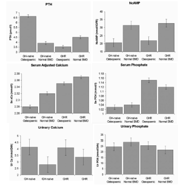
BONE REMODELING IN DRUG ADDICTS H. Wilczek*, J. J. Stepan Faculty of Medicine, Charles University, Prague, Czech Republic Aim. To study bone metabolism in asymptomatic heroin addicts and effect of methadone treatment. Methods. The study involved 37 drug addicts using heroin (mean dose 1.5 g / day), 31 men and 6 women (mean ± SD, age, 26 ± 5 yr, weight, 71 ± 10 kg, height, 180 ± 7 cm). The subjects were treated for one year with methadone (mean daily oral dose of 90 mg). BMD was measured using Hologic Delphi A. Serum aminoterminal propeptide of type I collagen (PINP) was measured by a radioimmunoassay (Orion Diagnostica). Serum type 1 collagen cross-linked C-telopeptide (S-beta CTX) was measured using the Elecsys (Roche). Results. At baseline, BMD at the lumbar spine and total femur was within normal mean range (T-score, -0.4 ± 0.8, and 0 ± 0.9, respectively), while at the femoral neck and distal forearm it was significantly decreased when compared with healthy adults, p < 0.01 (T-score, -0.7 ± 1, and 0.8 ± 0.8, respectively). Serum concentration of CTX (mean and 1 SD range, 814 and 543-1212 ng/l) and PINP (76, and 55-106 ug/l) was significantly increased as compared with healthy age and sex matched controls (p < 0.01). In men, serum concentration of testosterone (3.3, and 1.6 - 6.6 nmol/l) and estradiol (16 and 10-26 pmol/l) was significantly decreased. After one year of methadone treatment, serum CTX decreased significantly (p < 0.01); no significant change was observed in BMD. Conclusion. In the heroin addicts, bone remodeling is increased presumably as a result of effects of the drug on hypopituitary-hypothalamo-gonadal axis. THE RISK OF RAPID BONE LOSS IN INFLAMMATORY BOWEL DISEASE, MORE QUESTIONS THAN ANSWERS C. M. S. Schulte University Clinic of Essen, Germany Introduction: Patients with inflammatory bowel disease (IBD) are a 'risk population for osteoporosis eligible for preventive strategies'. Corticosteroids, inflammation and vitamin-D-deficiency are named risk factors. However, recent research has attenuated the dogma of IBD-associated osteoporosis. Age and body weight, but not IBD associated factors were main predictors of bone mass in Crohn's disease (CD) (1). In a population-based CD inception cohort, fracture risk was not elevated relative to controls (2). Long-term data in large populations are sparse (3). We perform a long-term analysis in a group of 250 IBD patients evaluating disease course and BMD every 18 months. Patients and methods: We analysed a subgroup of these IBD patients, where bone loss was not a mixture of senile, postmenopausal and IBD-associated bone loss by excluding the following patients from analysis: 1. Patients > 50 years 2. Postmenopausal women 3. Preexisting osteoporosis (T-score < - 2.5 SD) 4. Patients treated with bisphosphonates and/or fluoride. Annualized changes of bone mass (BMD measured by DEXA) were analysed in 94 IBD patients (32 ± 8 years; 68 CD, 26 ulcerative colitis; 47 male, 47 female), who were followed 36 ± 17 months. Average steroid during follow-up was 2.37 ± 4.7 mg/day. Patients were advised to take 1000 I.U. vitamin D and 1000 mg calcium daily. Results: Baseline BMD was normal at all sites: Z-score: spine: -0.27 ± 1.1, neck: -0.3 ± 1.2. Change of bone mass per year was small and positive: spine +0.55 ± 2.38, femoral neck +0.7 ± 4.2 %/year. Only few patients suffered from rapid bone loss > 2%/year (spine 3.3 %, femoral neck 11 % of the patients). Risk factors like type of disease, early disease onset, age, sex, BMI, steroid therapy and intestinal resection showed no correlation with the observed BMD changes. Discussion: Consecutive BMD measurements in 94 non-osteoporotic IBD patients indicated gain of bone mass. Only few patients showed rapid bone loss (> 2%/year) justifying an intervention with bisphosphonates in addition to the standard therapy (vitamin D + calcium during corticoid medication). Literature: (1) AJG 1999: 94 824 (2) Gastroenterology 2002: 123 468 (3) SJG 1999:34 696. PTH SENSITIVITY IS ALTERED IN OSTEOPAENIC PATIENTS WITH ADULT GROWTH HORMONE DEFICIENCY H. D. White1*, A. M. Ahmad1, K. Prabhakar1, S. Chandran1, B. H. Durham2, J. P. Vora1, W. D. Fraser2 1Department of Diabetes and Endocrinology, Royal Liverpool University Hospital, Liverpool, UK 2Department of Clinical Chemistry, Royal Liverpool University Hospital, Liverpool, UK PTH target-organ insensitivity may contribute to the reduced bone mineral density (BMD) associated with Adult GH Deficiency (AGHD). GH Replacement (GHR) leads to improvements in BMD and PTH target-organ sensitivity, but some patients remain osteopaenic despite full pituitary hormone replacement. BMD characteristics (normal/osteopaenic) in AGHD have not, as yet, been correlated with abnormalities in PTH sensitivity. Therefore, we investigated the difference in PTH and phosphocalcium metabolism in GH-naďve and GHR AGHD patients with normal and reduced BMD. (Mean femoral T-scores ± SEM are in brackets). 43 AGHD patients (25 GH-naďve patients (13 reduced BMD (-1.7 ±0.2), 12 normal BMD (1.0 ±0.3)), 18 GHR patients (10 reduced BMD (-1.4 ±0.3), 8 normal BMD (0.9 ±0.3))) were hospitalised for 24-hours. Half-hourly blood and 3-hourly urine samples were collected for PTH, calcium, phosphate and nephrogenous cAMP (NcAMP). All patients were adequately replaced on all other pituitary hormones. Results are shown in figure-1. Age, gender and disease duration did not differ significantly between the groups. In the GH-naďve patients, 24-hour mean PTH (p<0.001) and urinary calcium (p=0.03) were higher in the osteopaenic group, whilst NcAMP (p=0.004) and serum adjusted calcium (p<0.001) were lower. The GHR osteopaenic group had lower 24-hour mean PTH (p<0.001) but similar NcAMP than the GH-naďve osteopaenic group, with higher serum adjusted calcium and phosphate (p<0.001). PTH and phosphocalcium patterns were similar in GH-naďve and GHR patients with normal BMD. Our data demonstrates reduced target-organ PTH sensitivity in osteopaenic GH- naďve patients, which may contribute to the AGHD-associated reduction in BMD. Adequate parathyroid gland sensitivity to serum calcium probably exists in GHR osteopaenic patients given the appropriate PTH:adjusted calcium concentrations observed in this group. However, inappropriate NcAMP and serum phosphate levels suggest persistent renal PTH resistance in some patients which may result in the observed continued osteopaenic status.
PARATHYROID GLAND RESPONSITIVITY TO HYPO- AND HYPERCALCAEMIC STIMULI IN ADULT GROWTH HORMONE DEFICIENCY IS ALTERED FOLLOWING GROWTH HORMONE REPLACEMENT A. M. Ahmad1*, H. D. White1, J. Thomas1, M. T. Hopkins1, B. H. Durham2, J. P. Vora1, W. D. Fraser2 1Department of Diabetes and Endocrinology, Royal Liverpool University Hospital, Liverpool, UK 2Department of Clinical Chemistry, Royal Liverpool University Hospital, Liverpool, UK Adult GH deficiency (AGHD) is associated with osteoporosis. Alterations in parathyroid gland responsiveness to changes in calcium concentration play a role in the genesis of osteoporosis. We investigated PTH response to hypo- and hypercalcaemic stimuli in AGHD. 12 patients with severe AGHD were hospitalised, prior to GH replacement (GHR), at 0800h. Venous cannulae were inserted in each arm and half-hourly blood sampling commenced at 0900h and continued to 1800h. After 5 basal samples, sodium EDTA infusion was commenced at 1100h for 2h to induce hypocalcaemia in 6 patients (group 1). Hypercalcaemia was induced in 6 patients (group 2) by calcium gluconate over 2h. PTH, calcium and albumin were measured on all samples. GHR was commenced after baseline visit. The protocol was repeated at 3 and 12 months on GHR. In group 1, maximum PTH stimulation occurred at an adjusted calcium concentration (percentage drop) of 1.79mmol/L (23.8%) at 0 months. Following GHR, maximum PTH stimulation occurred at an adjusted calcium concentration of 1.92 (17.2%) at 3 months and 2.16 (12.9%) at 12 months (p<0.05). The maximum PTH response was a 365% rise before and 326% rise after 12 months (p=NS). In group 2, maximum PTH suppression occurred at an adjusted calcium concentration (percentage rise) of 2.79 (18.9%) at 0 months compared to 2.92 (16.7%) at 3 months (p=NS) and 2.84 (14.7%) at 12 months (p<0.01). The maximum PTH suppression was 75% before and 82% after 12 months (p=NS). The calcium set point (the calcium concentration at which the rate of PTH secretion is one half of its maximal value) progressively increased at 3 (p<0.01) and 12 months (p<0.001) in both groups following GHR. These results suggest increased parathyroid gland sensitivity to significantly smaller changes in serum calcium following GHR. Our findings may help explain the genesis of osteoporosis in AGHD patients. EFFECT OF GROWTH HORMONE REPLACEMENT ON PARATHYROID RESPONSIVENESS TO PARATHYROID HORMONE (1-34) INFUSION IN ADULT GROWTH HORMONE DEFICIENCY A. M. Ahmad1*, H. D. White1, J. Thomas1, M. T. Hopkins1, B. H. Durham2, W. D. Fraser2, J. P. Vora1 1Department of Diabetes and Endocrinology, Royal Liverpool University Hospital, Liverpool, UK 2Department of Clinical Chemistry, Royal Liverpool University Hospital, Liverpool, UK Alterations in both parathyroid gland sensitivity to changes in calcium concentration and end-organ response to the effects of PTH play a role in the development of osteoporosis. Since adult growth hormone deficiency (AGHD) is associated with increased prevalence of osteoporosis, we studied the effects of PTH (1-34) infusion on end-organ and parathyroid gland responsiveness. 6 patients with severe AGHD were recruited. All patients were admitted, prior to commencement of GH replacement (GHR), at 1300 h. Venous cannulae were inserted in each arm and half-hourly blood sampling was commenced at 1400 h. hPTH (1-34) was infused over 24 h. PTH, calcium and albumin were measured on all samples. GHR was commenced after the baseline visit. The protocol was repeated at 3 and 12 months on GHR. Following hPTH (1-34) infusion, adjusted calcium concentration increased after 10 h in untreated AGHD patients, whereas, after 12 months on GHR, adjusted calcium concentration increased within 5 h. After 12 months on GHR, the percentage increase at 4 h was 3.2 ±0.60% versus 0.83 ±0.66% in untreated AGHD patients (p<0.05) that remained significantly higher over the 24 h (17.8 ±2.9% versus 12.6 ±2.2%, p<0.05).There was no significant difference in the maximum PTH (1-84) suppression between visits. There was a significant increase in the calcium set-point (the calcium concentration at which the rate of PTH secretion is one half of its maximal value) after 3 (p<0.05) and 12 months (p<0.001) compared to baseline. We have demonstrated increased end-organ responsiveness to the effects of PTH resulting in a significant increase in calcium concentration in response to PTH (1-34) infusion, following GHR. together with the increase in calcium set-point these results may help understand the mechanisms underlying the genesis of osteoporosis in AGHD. DIFFERENT RESPONSES OF CORTICAL AND TRABECULAR BONE IN STEROID INDUCED OSTEOPOROSIS P. Augat*, S. Schorlemmer, G. Cotta, A. Ignatius, L. Claes University of Ulm, Ulm, Germany Trabecular bone is known to respond rapidly to steroid administration by a loss of bone mineral and a reduction of mechanical quality. However, little is known about steroid induced changes of mineral status and mechanical properties of cortical bone. The purpose of this study was to compare the responses of cortical and trabecular bone in an animal model of steroid induced osteoporosis. Sixteen ovariectomized female merino sheep were randomly assigned to steroid (Methylprednisolon 0.45 mg/kg BW) treatment for 6 months, or no treatment. Trabecular bone biopsies were harvested six and 12 months after the beginning of the study from the proximal tibia and the distal femur. The biopsies were scanned for apparent BMD by QCT, mechanically tested in compression, and analyzed by fluorochrome and conventional histology. Cortical bone was assessed after 12 months by QCT at the tibia, histology of tibia cross-sections, and mechanical testing of the entire metatarsus and bone specimens from the tibia. Steroid treatment reduced trabecular BMD by 27% (p<0.05) at six months and by 33% at 12 months. Also trabecular bone elastic modulus was markedly reduced by 36% at six months and by 62% at 12 months (p<0.05). In contrast neither the failure load of the metatarsus nor the elastic modulus of the bone specimens of the tibia differed between steroid treated and control animals. Cortical apparent BMD differed by less than 1.5% between steroid and control animals. Fluorochrome labeling demonstrated that steroid administration significantly turned down bone remodeling in trabecular bone as well as modeling and bone formation in cortical bone. After cessation of steroid administration periosteal and endosteal osteoid formation and osteonal remodeling returned to normal in cortical bone. However, in trabecular bone osteoid formation remained reduced by 53% and osteoid surface was still 70% smaller in steroid treated animals. Although six month of high dose steroid administration affected cortical and trabecular bone by massive reduction in bone modeling and remodeling, only trabecular bone suffered a significant loss of bone mineral and deterioration of mechanical properties. Furthermore, steroid appeared to remain active in trabecular bone for a longer time period than in cortical bone. BROADBAND ULTRASOUND ATTENUATION COMPARED WITH DUAL-ENERGY X-RAY ABSORPTIOMETRY IN SCREENING FOR OSTEOPOROSIS IN AN ASIAN POPULATION C. K. C. Chong*, L. Y. M. Lim, L. K. S. Lam Registrar, Changi General Hospital Registrar, Changi General Hospital Senior Consultant, Changi General Hospital Background: Quantitative ultrasound (QUS) of the calcaneus is an alternative technique to dual-energy x-ray absorptiometry (DEXA) for assessing bone mass. Various studies have demonstrated moderate correlation between broadband ultrasound attenuation (BUA) and bone density in the Caucasian population. Objective: To study the correlation between BUA and bone density in the Asian population, and its potential usage in screening for osteoporosis. Methods: 608 subjects had QUS examination of the calcaneus to establish the BUA normative data for the Singapore population. 32 of these subjects had DEXA scans of the hip done. Values for BUA of the calcaneus and bone mineral density of the hip are compared and correlated. Results: The correlation is good with a correlation coefficient of 0.6872 (p<0.0001). This is comparable to results of published series for the Caucasian population. Conclusions: QUS can be used as a screening tool for detecting osteoporosis in the elderly Asian population. ESTROGEN DEFICIENCY CAUSES BONE LOSS BY UPREGULATING T CELL PROLIFERATION AND LIFESPAN THROUGH IFNGAMMA INDUCED CLASS II TRANSACTIVATOR S. Cenci1,2, M. N. Weitzmann1, G. Toraldo1, C. Roggia1, Y. Gao1, W. P. Qian1, O. Sierra1, R. Pacifici1* 1Bone and Mineral Diseases, Washington University, St.Louis, MO, USA 2Molecular Immunology, San Raffaele Scientific Institute, Milano, Italy Expansion of the pool of TNFalpha-producing T cells in the bone marrow (BM) is critical for the bone wasting effect of estrogen deficiency, but the responsible mechanism is unknown. We report that ovariectomy (ovx) increases T cell number by enhancing T cell proliferation and lifespan through IFNgamma-induced class II transactivator (CIITA) in BM macrophages and T cells. Upregulation of CIITA in macrophages increases their MHCII expression and antigen presenting cell (APC) activity, leading to increased T cell activation and proliferation. Enhanced CIITA in T cells blunts activation-induced T cell apoptosis through repression of Fas ligand, leading to increased lifespan of active T cells. Upregulation of CIITA derives from increased production of IFNgamma by T helper-1 cells, resulting from increased secretion of IL-12 and IL-18 by BM macrophages. Ovx-induced T cell expansion and bone loss are prevented in vivo by both blockade of APC-induced T cell activation, and silencing of IFNgamma receptor signaling, which prevents IFNgamma-mediated induction of CIITA. Thus, increased IFNgamma-induced CIITA expression and the resulting enhanced T cell proliferation and lifespan are critical to the bone wasting effect of estrogen deficiency. SUBREGIONAL BONE MINERAL DENSITY MEASUREMENTS OF THE HAND IN PATIENTS WITH RHEUMATOID ARTHRITIS AND CONTROLS H. Franck*, J. Gottwalt, M. Welcker Center of Rheumatology, Oberammergau, Germany Patients with rheumatoid arthritis have bone loss to various degrees at different skeletal sides. The aim of the study was to evaluate whether subregional BMD of the hand is significant different in patients with rheumatoid arthritis and controls. Methods: BMD of the left hand was measured in the carpus and metacarpal joint II and III in 302 patients with rheumatoid arthritis and in 80 controls (ACR-criteria). Short-term measurements were performed in the same day and mid-term measurements after 1 month in 30 controls. Results: The short- term position errors were 0.9 - 1.4 %. Mid-term precision errors were 1.5 - 2.3 %. Patients with rheumatoid arthritis has significantly lower BMD values in the subregional carpus (0.405 ± 0.004 g/cm3) , metacarpal joint II (0.318 ± 0.036 g/cm3) and metacarpal joint III (0.326 ± 0.022 g/cm3) than controls (carpus: 0.424 ± 0.0045 g/cm3 versus metacarpal joint II 0.347 ± 0.036 versus metacarpal joint III 0.340 ± 0.037 g/cm3). There was a highly significant correlation between BMD total of the hand and the subregions (r = 0.92 - 0.98). Conclusion: Subregional measurements of the hand in patients with rheumatoid arthritis is the valuable tool assessing lower regional bone mass in patients with rheumatoid arthritis. Long-term studies are necessary to evaluate the importance of outcome parameters of subregional BMD in patients with rheumatoid arthritis. PERCUTANEOUS VERTEBROPLASTY IN THE TREATMENT OF VERTEBRAL FRACTURES P. Carpeggiani1*, M. Mazzantini2, V. Iacopetti2, O. Di Munno2 1Neuroradiology Unit, Ospedale S. Chiara, Pisa, Italy 2Rheumatology Unit, University of Pisa, Pisa, Italy Percutaneous vertebroplasty (PV) is a minimally invasive procedure consisting in the injection of polymethylmethacrilate (PMMA) into a vertebral body under radiological guidance. The use of this technique was first reported in 1984 for the treatment of painful spinal hemangiomas. Since then, its application was extended to stabilize pathologic fractures due to spinal metastasis and finally to treat vertebral osteoporotic fractures (VOFs). The latter is now becoming the principal indication of VP given the increasing number of patients with clinical VOFs, who are frequently refractory to traditional treatment, including bed rest, bracing and analgesics. PV, which is performed under local anesthesia, provides a significant pain relief within the first few days after the procedure and a mechanical restoration of the vertebrae weakened by the disease. The risk of fracture of osteoporotic vertebrae adjacent to that injected with hard PMMA has not been shown to be increased in a multicenter trial. In our Center 50 patients (age range 23-88 years) have been treated with VP: 4 affected with spinal hemangioma, 6 with metastases, and 40 with VOFs. In such patients 75 procedures have been performed, 25 on thoracic and 50 on lumbar vertebrae (mean number of vertebrae treated for each procedure 1.5; range 1-3). Patients with VOFs are being enrolled in a longitudinal study aimed to assessing the long-term outcome of VP. Before the procedure and after 1, 3, 6 and every 6 months thereafter this follow-up study will evaluate spinal pain by Visual Analogic Scale (VAS); functional impairment by self administered Health Assessment Questionnaire (HAQ) and quality of life by Quality of Life Questionnaire of the European Foundation for Osteoporosis (QUALEFFO); and the occurrence of new vertebral fractures by annual x-ray examination of thoracic and lumbar spine. The mean follow- up period of those patients is currently 9 months (range 1-14). Our preliminary data showed that in all patients VAS, HAQ and QUALEFFO scores improved significantly with respect to baseline values. During VP procedure no serious adverse event occurred, with the exception of an asymptomatic pulmonary microembolisation due to venous likeage of PMMA BONE MINERAL DENSITY AND BODY COMPOSITION IN NERVOUS ANOREXIA BEFORE AND AFTER A NUTRITIONAL REHABILITATION PROGRAM D. Gatti1*, S. Adami1, V. Braga1, F. Colapietro1, R. Prizzi1, M. Rossini1, T. Todesco2, R. Dalle Grave2 1Rheumatological Rehabilitation, University Hospital Valeggio S/M, Italy 2Department of Nutritional Rehabilitation, Villa Garda, Peschiera del G., Verona, Italy Osteoporosis is a frequent complication of alimentary disorders. An example are young women with nervous anorexia (NA) in whom bone mineral density (BMD) is severely decreased. Little is known on the relative contribution to osteopenia of fat and lean mass and on the effect on BMD of an effective nutritional rehabilitation program. We studied 57 women (aged 25.8, 6.7SD years) with NA (mean BMI: 14.8, 2,0SD) and 27 young healthy control women with BMI < 23 (mean 20.1, 1.9SD) in whom BMD (spine and hip) and body composition (total body ) were assessed by Hologic QDR 4500. In NA patients BMD were significantly lower than in controls both at the spine (T-score: -2.0, 1.9SD) and the hip (T-score: -1.7, 0.9SD). The BMDs values were significantly related to lean mass but not fat mass. Forty of the NA patients underwent a residential program of nutritional rehabilitation over 3 months. Eighteen of the control women agreed to follow a diet containing an amount of calcium and vitamin D identical to that of NA patients. At the end of the nutritional program body weight rose by 12.5, 4.9SD Kg in the NA patients. This was associated with increases of BMD by 2.4, 4.4SD% (p<0.001) vs. baseline and changes in controls at the hip and by 0.9, 3.7SD (Not Significant) at the spine . The BMD changes both at the hip and the spine were significantly (p=0.01) related to the changes of lean mass but not of fat mass. In conclusion in patients with NA the BMD at the spine and hip is 20% lower than in control subjects. A successful nutritional rehabilitation program increases BMD within 3 months, despite persistent amenorrhea, and these increases are strictly related to changes in muscle mass rather than fat mass. DURABILITY OF THE HUMERAL BONE OF WHITE RATS AT REPARATIVE REGENERATION OF THE TIBIAL BONE V. I. Luzin*, L. V. Stklyanina, D. S. Belushchenko, D. V. Mochalov State Medical University, Lugansk, Ukraine Research is carried out on 36 white female rats by initial weight 130-150 grams. 18 animals under aether narcosis a standard pine forest in diameter of 2,2 millimeters rendered foraminous through defect on border of proximal metaphysis and diaphysis of both tibial bones. As a control group served 18 intact rats. All animals contained in standard conditions of vivarium. Later 10, 30 and 90 days after drawing defect of animals were took out from experiment according to rules evtanasia under an aether narcosis. For research humeral bones are allocated which durability investigated at a bend with a speed of loading 0,25 millimeters in minute before destruction. Expected the sag, the breaking point, the module of elasticity and the minimal work of destruction. At intact animals during supervision with increase in age value of a humeral bone sag decreased, and values of the breaking point, the module of elasticity and the minimal work of destruction grew. Have established, that by 10 day after drawing defect in tibial bones the tendency to decrease in durability of a humeral bone was outlined: value decreased for 18,95% sag and on 6,02% the minimal work of destruction of a bone went down. In 30 days reduction in durability of a humeral bone was maximal: breaking point was on 19,53% less than control values, and work of destruction on 25,70%. Thus value of sag increased for 20,87%. As value of the module of elasticity authentically did not change, it is necessary to assume presence of qualitative changes in organic matrix which condition last both parameters characterize. Such changes testify about increase in fragility of a humeral bone and are characteristic, on our data, for osteopenic or osteoporotic conditions. By 90 day the revealed changes substantially were levelled. Thus, in conditions of actively current processes reparative regenerations in tibial bones of white rats of reproductive age mechanical durability of bones of other segments of a skeleton (namely a humeral bone) decreases. Probably, it is connected by that in conditions of presence of the center of reparative regenerations in bone system as a whole develop displays expression of the osteopenia condition. OSTEONECROSIS IN LONG TERM SURVIVORS AFTER BONE MARROW TRANSPLANTATION C. M. S. Schulte1*, D. W. Beelen2 1Department of Internal Medicine, University Clinics of Essen, Germany 2Department of Bone Marrow Transplantation, University Clinics of Essen, Germany Introduction: Organ transplantation is associated with rapid bone loss and harmful complications like fractures and osteonecrosis. Long term data clarifying the situation in patients after bone marrow transplantation are sparse. To illuminate the problem, we continuously follow disease course and BMD in a group of 300 BMT patients yearly since 1996. Aim: To quantify occurrence of osteonecrosis after BMT and to identify risk factors. Patients and methods: We performed a subanalysis of 119 long-term survivors (survival > 1000 days). Mean follow-up was 4.3 ± 1 years. BMD was measured by DEXA (Lunar DPX-L). X-ray or MRI was not performed routinely, but when patients complained pain. Patients were advised to take 1000 I.U. vitamin D plus 1000 mg calcium daily. Patients showing an osteoporotic BMD (T-score < -2.5 SD) were treated with bone active therapy according to the recommendations for postmenopausal osteoporosis (mainly bisphosphonates) and excluded from further analysis. Univariate Kaplan-Meier-statistic was performed to evaluate the influence of these potential risk factors: age at transplantation, gender, steroids applied after BMT, baseline BMD, amount/temporal sequence of bone loss after BMT. Results: Spinal BMD was normal before BMT (T-score -0.19, Z score -0.25). Steroid intake was 30 ± 25 mg/day during first post-tx-year, 10 ± 12 during further follow-up. First post-tx-year bone loss was rapid (5.4 ± 6.6%/year), while there was regain of bone mass during further follow-up. 24 patients (20%) started with bone active medication 2 ± 1.5 years after BMT. 15 patients (13 %) developed an osteonecrosis 2.6 ± 1.5 years after BMT. Altogether 18 hip, 4 knee and one talus osteonecrosis with multiple events in 5 patients were observed. Rapid bone loss within the first year was the risk factor correlating best with the occurrence of an osteonecrosis (Wilcoxon-test: 0.0002). All other risk factors, including BMD at baseline and steroid intake, were of minor or no influence. Discussion: Osteonecrosis is a major and frequent complication after BMT. Rapid bone loss within the first post- tx-year is the most important risk factor. These data indicate that early, rapid bone loss after BMT is devastating independent from potential recovery of bone mass during further follow-up. ASSOCIATION OF VITAMIN D RECEPTOR GENE POLYMORPHISMS (BSMI AND FOKI) WITH BONE DENSITY AND OSTEOPOROTIC FRACTURES: A CASE CONTROL STUDY A. Rogers*, F. Gossiel, J. A. Clowes, R. Eastell Bone Metabolism Group, University of Sheffield, UK Osteoporosis has a strong genetic component and vitamin D is essential for normal bone metabolism. Polymorphisms of the vitamin D receptor (VDR) gene have thus been studied extensively in relation to bone mass. However there have been fewer studies powered to detect associations between VDR genotype and osteoporotic fracture. The aim of this study was to examine the relationship between BsmI and FokI VDR polymorphisms and osteoporotic fracture. We studied 719 postmenopausal women ages 55 to 80 (mean 68) years. Of these, 265 women had sustained either a distal forearm (79), humeral (69), vertebral (66) or hip (51) fracture. The remainder were a population-based sample of 454 postmenopausal women from the Sheffield centre of the osteoporosis and ultrasound study (OPUS) study. Bone mineral density (BMD) was measured by DXA (Hologic QDR 4500A) at the lumbar spine (LSBMD) and total hip (THBMD). BsmI and FokI polymorphisms were assayed using an ABI Prism 7200 Sequence Detection System (Taqman). After correction for multiple comparisons, P < 0.008 was significant at the 95% confidence interval. LSBMD was significantly lower (P<0.0001 ANOVA) in the fracture cohort (Mean 0.794; (SEM 0.157) g/cm2) compared to the population-based cohort (1.054 (0.187) g/cm2). THBMD was significantly lower (P<0.0001) in the fracture cohort (0.752 (0.139) g/cm2) compared to the population-based cohort (0.906 (0.145) g/cm2). There was no difference in the frequency of BsmI and FokI genotypes between fracture and control groups (P > 0.05, ChiSq analysis). Fracture, age and weight, but not genotypes were significant factors in relation to LSBMD and THBMD in the whole group by multiple regression analysis. Haplotype analysis demonstrated that the bbFF haplotype had significantly higher LSBMD when fracture subgroups (upper limb, vertebral and hip) were analysed (All P < 0.004). There was a similar trend for THBMD, but this did not reach significance after correction for multiple comparisons (All P<0.05). BsmI and FokI polymorphisms, individually or in combination, did not predict fracture independent of BMD by logistic regression. In conclusion VDR polymorphisms are not related to osteoporotic fracture in this cohort of elderly women. However our results suggest that individuals with the bbFF haplotype may have a higher bone density in the region of the lumbar spine. POLYMORPHISMS IN THE TRANSFORMING GROWTH FACTOR BETA1 GENE AND PERIMENOPAUSAL BONE MASS, EARLY POSTMENOPAUSAL BONE LOSS AND RESPONSE TO HORMONE REPLACEMENT TREATMENT B. L. Langdahl1*, B. Abrahamsen2, P. Vestergaard1,2, C. L. Tofteng2, N. Kolthoff2, J. S. Madsen3, L. Mosekilde1,2 1Department of Endocrinology, Aarhus Amtssygehus, Denmark 2Danish Osteoporosis Prevention Study Group, Denmark 3Department of Clinical Biochemistry, Odense University Hospital, Denmark Transforming Growth Factor beta1 is the most abundant growth factor in bone and plays an important role in the coupling of bone resorption and bone formation. We have previously shown that three polymorphisms in the TGF-beta1 gene: C-1348-T, T29-C and T861-20-C are associated with reduced bone mass and increased risk of osteoporotic fractures. We therefore wanted to investigate if these effects are mediated through peak bone mass, early postmenopausal bone loss or the response to HRT after menopause. We determined the genotypes of the three polymorphisms in 1684 women participating in the DOPS. The women were within two years of menopause at baseline and 35 % were subsequently treated with HRT. We have previously found the TT-genotype of the T861-20-C polymorphism to be associated with reduced bone mass and increased fracture risk in an older population. However, in these perimenopausal women, we found that women with the variant genotypes (CT and TT) had lower BMD (g/cm2) compared with women with the wildtype genotype (CC) at all measured sites (see table). The C-1348-T and T29-C polymorphisms are in strong linkage disequilibrium. BMD was higher at all measured sites in women with the variant genotypes, however the difference did not reach significance. These results are in agreement with our previous study. The women were followed for 5 years and bone loss was significantly affected at the total hip by T861-20C: Women with the TT genotype lost 5.45±4.69 % compared with 3.75±4.82 % in women with the CT genotype and 4.37±4.50 % in women with the CC genotype (p<0.01). At the femoral neck bone loss was greater in women with the TT genotype in position -1348 and CC genotype in position 29. These effects were abolished by HRT. In conclusion: We have demonstrated that common polymorphisms in the TGF- beta1 gene are associated with reduced perimenopausal BMD and increased bone loss in early postmenopausal women.
RELATIONSHIP BETWEEN VITAMIN D BINDING PROTEIN GENE POLYMORPHISMS AND BONE MINERAL DENSITY OF LUMBAR SPINE AND FEMORAL NECK IN POSTMENOPAUSAL RUSSIAN WOMEN M. Yu Krylov*, V. A. Maykotkin, L. I. Benevolenskay Institute of Rheumatology Russian Academy of Medical Sciencies, Moscow, Russia The name of vitamin D binding protein (DBP) is derived from its ability to bind the principal vitamin D metabolites:25-hydroxyvitamin D and 1.25 -dihydroxyvitamin D the major key metabolites of bone turnover. DBP may also play an important role in osteoclast differentiation. There are two known polymorphisms in exon 11 of the DBP gene that result in amino acid variants: at codons 416 GAT-->GAG (Asp-->Glu) and 420 ACG-->AAG (Thr-->Lys). The aim of this study was to investigate whether DBP gene polymorphisms are associated with bone mineral density (BMD) of the lumbar spine and femoral neck in postmenopausal Russian women. We studied 70 unrelated postmenopausal women (mediana age 71, ages 60-79 years) with osteoporosis (OP) without medication and 51 control women (mediana age 69, ages 58-81 years). BMD at the lumbar spine and femoral neck were measured by dual X-ray absorptiometry (Hologic 4500). DNA was isolated from blood and variations at codons 416 and 420 were examined by polymerase chain reaction-restriction fragment length polymorphism (PCR-RFLP). The was no apparent effect of DBP phenotypes on BMD in control group, but there was a relationship between certain DBP genotypes and reduced BMD in women with OP. Femoral neck BMD was significantly lower in women with SS genotype than 2F genotype (0.606 ±0.01 SEM g/cm2 and 0.654 ±0.02 SEM g/cm2 respectively, p<0.05) and lumbar spine BMD was lower in patients with genotype FS than SS genotype (0.674 ±0.02 SEM g/cm2 and 0.757 ±0.04 SEM g/cm2 respectively,p=0.056).In conclusion our results suggest that polymorphism in exon 11 of the DBP gene may have an important effect on bone density in postmenopausal women with osteoporosis. LACK OF ASSOCIATION OF TRANSFORMING GROWTH FACTOR- BETA1 T29->C GENE POLYMORPHISM WITH CALCANEAL QUANTITATIVE ULTRASOUND PARAMETERS AND PLASMA TGF- BETA1 LEVELS IN POSTMENOPAUSAL KOREAN WOMEN J-M. Koh1, S-W. Kim2, Y-E. Chung3, J. S. Hong4, S-Y. Kim5, G. S. Kim1* 1Division of Endocrinology and Metabolism, University of Ulsan College of Medicine, Asan Medical Center, Seoul, Republic of Korea 2Department of Internal Medicine, Incheon Sarang Hospital, Incheon, Republic of Korea 3Department of Internal Medicine, Seoul Veterans Hospital, Seoul, Republic of Korea 4Asan Life Science Institute, Seoul, Republic of Korea 5Department of Orthopedic Surgery, Kyungpook National University, Taegu, Republic of Korea Transforming Growth Factor-beta1 (TGF-beta1) may be a pivotal role in the bone- remodeling process. Recently, it has been reported that a T->C polymoprhism at nucleotide 29 of the signal sequence of the TGF-beta1 gene is associated with bone mineral density at the lumbar spine and serum TGF-beta1 levels in postmenopausal Japanese women. However, the controversial result has been reported for this association in postmenopausal German women. We studies the possible association of the TGF-beta1 gene T->C polymorphism with quantitative ultrasonic parameters and with plasma TGF-beta1 levels in postmenopausal Korean women. Broad ultrasound attenuation (BUA) and speed of sound (SOS) were measured by quantitative ultrasound at the calcaneus in 211 healthy postmenopausal women aged 60 to 69 years who were part of the population-based Jungup osteoporosis prevalence study. Genotyping for polymorphism of TGF-beta1 gene was performed using polymerase chain reaction methods. The frequencies of the TT, TC and CC genotypes in the study population were 32.3%, 42.7% and 25.1% respectively. Calcaneus BUA in individuals with the TT genotype did not differ from those with TC or CC genotype (95.2 ±8.7 vs. 94.5 ±8.7 vs. 95.2 ±11.4, p = 0.884). Neither SOS nor SI value also showed difference among the TGF-beta1 genotypes. Among the study subjects, 96 (45.5%) were within normal range of SI values, 97 (46.0%) were osteopenic, and 18 (8.5%) were osteoporotic. The distribution of TGF-beta1 genotypes was not different between normal, osteopenia, and osteoporosis women (c2 = 0.258, p = 0.611). Furthermore, the plasma concentration of TGF-beta1 in study subjects did not show any difference among the genotypes. In conclusion, our study demonstrated that T29->C gene polymorphism was associated neither with quantitative ultrasound parameters nor with plasma TGF-beta1 levels in postmenopausal Korean women. THE PREVALENCE OF SECONDARY CAUSES OF OSTEOPOROSIS IN WOMEN WITH ASTMA M. L. Menshikov1*, L. V. Menshikova1,2 1Institute for Advanced Medical Studies, Irkutsk, Russia 2Regional Rheumatological Department, Irkutsk, Russia The purpose of this study was to determine the prevalence of secondary causes of osteoporosis (OP) in postmenopausal women with asthma. The study comprised 61 postmenopausal women with mean age 59,5 (7,4) y.o. (range 47-74) with asthma using continuous oral corticosteroids (CS), mean dose consist of 9 mg/day and 33 patients without CS (mean age 57,2 y.o.). Control group consisted of 326 females, matched with respect to age, body weight and body mass index, menopausal status. BMD was measured by DEXA (DPX-IQ, Lunar) at lumbar spine (L2-L4) and hip in the all group. Biochemical markers of bone turnover (serum osteocalcin, bone alkaline phosphatase, urinary deoxypiridinoline, beta-crosslaps) were measured. According to the DEXA parameters among patients without CS osteopenia and OP were observed in 24,2% and 18,2% at the lumbar spine, and in 45,4% and 9% at the femoral neck respectively. The patients with CS had consistently the lowest BMD. OP was prevalent at the lumbar spine 52,5% than at femoral neck-31,1%, osteopenia had 29,5% and 52,5%. T-score = -2,6 SD and 1,8 SD; mean BMD was 0,829(0,161) g/cm2 and 0,767(0,133) g/cm2 respectively. Fractures in minimal trauma had 37(60,7%) women using CS. Mean age menopause was 46,8 y.o. and less than in the control group and in the women without CS. We find negative correlation with postmenopause age, cumulative prednisolone dose, low body mass index. There were at the same time the increased markers of bone resorption, decreased markers of bone formation. Conclusions: The results of this study can suggest that, prolonged oral CS therapy increases the risk of secondary osteooposis as well as osteopenia and fractures. RELATION OF GENOTYPES OF LACTASE HYDROLASE, VITAMIN D AND ESTROGEN RECEPTOR AND COLLAGEN I ALPHA 1 GENES TO BONE MINERAL DENSITY AND BONE FRACTURE INCIDENCE IN POSTMENOPAUSAL WOMEN B. M. Obermayer-Pietsch1*, C. M. Bonelli1, D. E. Walter1, A. Fahrleitner-Pammer1, A. Berghold2, W. Goessler3, V. Stepan1, H. Dobnig1, G. Leb1, W. Renner4 1Dep. of Internal Medicine, Karl-Franzens-University, Graz, Austria 2Insitute for Medical Informatics, Statistics and Documentation, Karl-Franzens- University, Graz, Austria 3Institute of Chemistry - Analytical Chemistry, Karl-Franzens-University, Graz, Austria 4Institute of Molecularbiological Analytics, Graz, Austria Background Many polymorphic variations in bone-related genes have been investigated for their association with bone mineral density and bone fractures. We recently reported on a -13910 T/C dimorphism nearby the lactase-phlorizin hydrolase (LCT) gene, strongly associated with adult lactase non persistence. This polymorphism was tested together with genotypes of the vitamin D receptor (VDR, BsmI and FokI), the estrogen receptor (ER, XbaI and PvuII) and the collagen I alpha 1 genes (COLIA, Sp1) to evaluate their influence on bone mineral density (BMD) and the incidence of bone fractures in postmenopausal women. Methods We determined LCT, VDR, ER and COLIA genotypes in 224 postmenopausal women using polymerase-chain-reaction-based assays. All genotypes were related to age-adjusted BMD values (z-scores) at the lumbar spine and the femoral neck (DEXA, Hologic 4500 plus), and the occurrence of nonvertebral fractures using multivariate methods. Results We found a significant association between LCT genotypes and BMD either at the lumbar spine (p=0.004) and all sites of the femoral neck (p=0.006). LCT genotypes were the only ones to contribute significantly to BMD, explaining 2.8 - 2.9% of age- adjusted BMD values and 2.1% of bone fracture incidence. VDR but not ER or COLIA genotypes modified BMD values in LCT genotype groups by further significantly decreasing BMD z-scores at all sites measured in LCT CC (lactase deficient) women with additional VDR BB or FF genotypes. Conclusions About 3% of lumbar and femoral BMD and consecutive bone fractures can be explained by LCT(T/C-13910) polymorphisms, whereas variations in other candidate genes may have less influence on BMD and bone fractures. However, we suppose additive effects in genotypes of calciotropic genes. LCT variations are very common in the European population, and genetic testing for lactose intolerance may help to define persons at risk for osteoporosis and bone fractures. VERTEBRAL DEFORMITY ASSESSMENT BY MORPHOMETRIC X- RAY ABSORPTIOMETRY (MXA) V. Baldini*, D. Diacinti, E. D'Erasmo, M. Celi, C. Letizia, E. Tomei, S. Minisola, G. F. Mazzuoli University 'La Sapienza', Rome, Italy The purpose of this study was to compare MXA and morphometric radiography (MRX) of the spine for the identification of vertebral deformities in a group of postmenopausal women of Rome. Materials and methods: on 300 postmenopausal women of Rome (ages 45-80 years) referred to our Osteoporotic Centre, were acquired lateral images of the spine by conventional x-ray tube and by dual energy X- ray absorptiometry (DXA) using the Hologic QDR-4500 densitometer (Hologic, USA). On these images one radiologist (D.D.) performed the vertebral morphometry using a specified software to measure the vertebral bodies heights from T4 to L4. The vertebra was considered fractured if any of the three heights ratios was <3SD compared to the respective normal mean values of fertile women (Mc Closkey). For gold standard identification of vertebral fractures we used a consensus reading of Genant's semiquantitative (SQ) method by an experienced radiologist (E.T.) and one of the authors (E.D.) Results: MXA categorized 26.3% of the 300 subjects as having one or more vertebral fractures (sensitivity 93%; specificity 99%) whereas MRX identified 26.7% as having fracture (sensitivity 94%; specificity 99%). MXA and MRX agreed that 200 subjects were normal and 79 subjects were fractured ( k=0.79- 0.76). MXA identified 86% of the 3900 vertebrae as normal, 10% as deformed, and 4% were not visualized sufficiently for analysis, whereas MRX identified 88%, 11,7% and 0.3% of these vertebrae as normal deformed, or unanalyzable, respectively. Conclusion : the two morphometric methods, MRX and MXA, disagree particularly to identify mild deformities in the upper thoracic spine because poor MXA image quality. Nevertheless this disadvantage of MXA has poor clinical relevance because of the lower prevalence of osteoporotic vertebral fractures in levels T4-T6. In fact in this study only subject was incorrectly classified as normal by MXA. A SPECIFIC MUTATION IN THE P2X7 RECEPTOR DNA SEQUENCE MAY BE RESPONSIBLE FOR IMPAIRED BONE QUALITY S. Ohlendorff1*, M. Friberg-Hitz2, C. L. Tofteng1, J. E. Beck Jensen1, N. R. Jřrgensen1 1The Osteoporosis and Bone Metabolic Unit, Department of Endocrinology, Copenhagen University Hospital H:S, Hvidovre, Denmark 2The Bone Laboratory, Roskilde County Hospital, Koege, Denmark P2X7 is a purinergic receptor inducing a pore responsible for apoptosis in osteoclasts. A single nucleotide polymorphism for P2X7 has recently been discovered. P2X7 receptors seem to induce apoptosis in osteoclasts, and impaired function of this receptor might therefore be responsible for decreased apoptosis in osteoclasts, increased bone resorption and consequently responsible for excessive loss of bone. The aim of this study was to investigate the distribution of a specific mutation in two Danish cohorts differing in bone health status. The polymorphism was examined using PCR-RFLP and sequencing. Two subject groups were included for comparison of allele frequencies of the mutation. Group one comprise of 52 osteoporotic postmenopausal women with osteoporotic fractures, hospitalized at Roskilde County Hospital Koege, Denmark. Group two comprise of 137 postmenopausal women recruited from the Danish Osteoporosis Prevention Study (DOPS), at the Osteoporosis Research Unit, Copenhagen University Hospital Hvidovre, Denmark, and is considered to represent the background population. The 189 samples analysed, revealed a strikingly different genotype distribution in the two groups. The osteoporotic fracture group comprised 30 wildtype subjects, 19 heterozygous subjects and 3 homozygous subjects. The normal control group comprised 110 wildtype subjects, 20 heterozygous subjects and 7 homozygous subjects. This gives a mutant allele frequency of 0.240 for the osteoporotic fracture group and 0.124 for the normal control group. The mutant distribution between the groups is significantly different with a p-value = 0.0036, as assessed by the Fishers Exact Test. The increased allele frequency found for the osteoporotic group compared to the background control group, suggests that the specific mutation could be associated with pathogenesis of osteoporosis and therefore be responsible for increased risk of osteoporotic fractures. Thus, the mutation might be used as a marker of bone quality.
DOES OBESITY ACCELERATE LATE POSTMENOPAUSAL FEMORAL BONE LOSS? R. J. Saarelainen1,2*, R. J. Honkanen1, M. Tuppurainen3, L. Niskanen2 1Research Institute of Public Health, University of Kuopio, Kuopio, Finland 2Department of Medicine, Kuopio University Hospital, Kuopio, Finland 3Department of Gynecology, Kuopio University Hospital, Kuopio, Finland Overweight is associated with decreased postmenopausal bone loss. We examined the association of obesity and BMD change further by relating it to time since menopause and bone site in 398 early postmenopausal women from the random sample of the population-based OSTPRE cohort. Bone mineral density (BMD) was measured with DXA (Lunar DPX) at the spine (L2-L4) and hip (femoral neck)) three times: at baseline, at 5.9-year and at 10.5 year- follow-up. The study population was formed by excluding women who were premenopausal at 5.9-year follow-up, women without information about menopause, women who had used HRT, bisphosphonates or calcitonin during follow-up and women who lacked a complete series of valid spine or hip BMD results. The mean follow-up times were 5.9 (range 3.9-8.0) for the first and 4.6 (range 2.9-7.6) years for the second study period. We divided the study population into three groups: PERI=menopause during the first study period (N=101), EARLY= postmenopausal<=5 years at baseline (N=177) and LATE=postmenopausal>5 years at baseline (N=120). The mean baseline age was 53.8 (SD 2.8, range 49-59) years for the total study population, and 51.2 (1.9), 53.7 (2.5) and 56.1 (1.8) years for the PERI, EARLY and LATE groups, respectively. Mean baseline height, weight and body mass index (BMI)were 161.4 (5.06) cm, 71.3 (11.9) kg and 27.4 (4.4)kg/m2. BMI was categorized as follows: <25 (N=127), 25-29.9 (N=182) and 30+ (N=89). Mean annual BMD changes during the 1st and 2nd study periods were: -0.6 and +0.03 % for spinal and -0.7 and -0.8 % for the femoral BMD, respectively. These changes were -0.8 and -0.2 % for spinal and -0.7 and -0.6 % for femoral in normal- weighted (BMI<25) but -0.3 and +0.1 % for spinal (p=0.018, paired t-test) and -0.7 and -1.2 % for femoral (p=0.008) BMD changes in obese (BMI>30) women, respectively. In obese LATE postmenopausal group (N=23) these changes were: +0.2 and +0.4% (ns) for spinal and -0.4 and -1.1 % (p=0.02) for femoral BMD change, respectively. Femoral bone loss rate seems to accelerate in late postmenopause of obese women. PREVALENCE OF OSTEOPENIA AND OSTEOPOROSIS IN 11975 POSTMENOPAUSAL ARGENTINE WOMEN F. Massari*, D. Bron, M. Lopez, S. Sanchez, M. Toccheton, L. Wasserman, B. Zanchetta, J. R. Zanchetta IDIM: Metabolic Research Institute, Bs.As., Argentina Taking into consideration the methodological limitations of the prevalence studies and the lack of information of this issue in our country, we describe the prevalence of Osteopenia/Osteoporosis in postmenopausal women (50 years old or older) participating in a free detection survey with Lumbar Spine (LS) and Femoral Neck (FN) DEXA (OMS Criteria), taking as diagnostic the lowest density region). After the inclusion/exclusion criteria, were selected 11975 women, mean age: 67 years (50-94), mean menoage: 48 years, and mean BMI: 28.8 Kg/mt2. Osteopenia prevalence was 65.46 % (n: 7839); of that, 27.9 % was mild (TS: -1/- 1.49), 38.3 % was moderate (TS: -1.5/-1.99), and 33.8 % was severe (TS: -2/-2.49). Osteoporosis prevalence was 15.91 % (n: 1905), and the discrimination by diagnostic region, showed that 52.23 % (n: 995) was only affected at LS, 17.27 % (n: 329) only at FN, and 30.5 % (n: 581) at both regions. This pattern presented some differences between decades of age, but confirm the diagnostic utility of FN in women younger than 65 years old, and for the LS in older women, even after 80 years old. The prevalence of self-reported clinical fractures (wrist, hip, vertebral) was similar between osteoporosis and severe osteopenia patients (29.73% vs 25.36% p: 0.268) In conclusion; we found some differences between our prevalence of osteopenia/osteoporosis and the findings of the references studies (probably due to multiple factors; technological, racial, geographic, etc). We reinforce the convenience of the diagnostic evaluation of both densitometric regions (LS/FN) at all ages. We remark the potential mistake of underestimate the fracture risk of severe osteopenia THE ANALYSIS OF BIOCHEMICAL BONE MARKERS (OC, ALP, NTX, DPD) DEPENDING ON THE MENSTRUATION CYCLE AND EATING ATTITUDE OF BALLET DANCERS D. H. Kim1, H. Y. Lee1*, Y. S. Kim1, Y. R. Kim2, Y. B. Yoon3 1Department of Physical Education, Chonnam National University, Gwangju, Korea 2Daebul University, Jeonnam, Korea 3Chonnnam Techno College, Jeonnam, Korea This study consists of 159 subjects, 45 professional dancers, and 114 student dancers with abnormal and normal eating and menstruation disorders. Eating and menstruation disorders were investigated depending on the presence and absence of eating and menstruation by EAT-26 and menstrual self report. The subjects were divided into two groups : normal and abnormal of eating and menstruation. A study of their effects was conducted on biochemical bone markers(OC, ALP, NTx, DPD). The investigated results of biochemical bone markers were as follows; When comparing among groups the biochemical bone markers, which depends on the presence and absence of eating and menstruation disorders, ALP in the professional dancers was significantly lower than in the student group. ALP in the abnormal menstruation group was significantly lower than in the normal menstruation group. There was also significant interaction between the groups and menstruation. The relationship of the biochemical bone markers which depends on the presence and absence of menstruation, showed a positive relationship between ALP and NTx in the normal menstruation group. There was a positive relationship between OC and ALP in the abnormal menstruation group. THE EFFECT OF STANDARD HEPARIN AND TWO LOW- MOLECULAR-WEIGHT HEPARINS ON MECHANICAL PROPERTIES OF THE FEMUR IN RATS J. Folwarczna*, W. Janiec, I. Kaczmarczyk-Sedlak, M. Pytlik, L. Sliwiński, U. Cegiela, B. Nowińska Department of Pharmacology, Medical University of Silesia, Sosnowiec, Poland Long-term administration of heparin is associated with development of osteopenia in humans and experimental animals. Low-molecular-weight heparins have been reported to cause weaker unfavourable effects on the histomorphometric parameters of the rat skeletal system than standard heparin. The present study was carried out to compare the effects of administration of standard heparin and two low-molecular- weight heparins (nadroparin and enoxaparin) on bone mechanical properties in rats. The experiment was conducted on 3-month-old female Wistar rats. Heparin was administered in doses of 1000 or 2000 IU/kg s.c. daily, nadroparin and enoxaparin were administered in doses of 1000 or 2000 anti-Xa IU/kg s.c. daily, for 28 days. Bone mass, length, diameter, mineral content (in the tibia, femur and L-4 vertebra) and mechanical properties of the whole femur (extrinsic stiffness, ultimate and breaking load, deformation caused by the applied load) and the femoral neck (load at fracture) were examined. Administration of the investigated heparins did not affect the macrometric parameters of bones, but caused slight decreases in the bone mineral content/bone mass ratios in comparison with the control group. The changes were noticeable the most after administration of standard heparin in a dose of 2000 IU/kg s.c. daily. Administration of standard heparin, nadroparin and enoxaparin induced unfavourable changes in mechanical properties of the femur. The investigated heparins decreased the breaking load of the whole femur (the load applied at fracture). Contrary to the control group, in which the values of ultimate load and breaking load were close to each other, after administration of all the investigated heparins (especially in higher doses) the breaking load was much less than the ultimate load, indicating development of changes in the bone structure. Only standard heparin administered in a dose of 2000 IU/kg s.c. daily weakened the femoral neck. OSTEOPOROSIS IN TYPE 2 DIABETICS K. M. Prasanna Kumar*, M. Dharmalingam, S. Karthik Shankar, K. Geetha Bhat M.S.Ramaiah Medical College, India Introduction: Diabetes of long standing duration may affect the bones and lead to osteoporosis.Review of literature showed conflicting results from normal 'bone density' to decreased 'bone density' in diabetics compared to non-diabetics. Aim: To study the impact of diabetes on bone mineral density(BMD) and prevalence of osteoporosis in diabetic SouthIndian men and women. Method and Materials: 40 diabetic and 58 non diabetic men, 57 diabetic and 191 non diabetic women in the age group of 51 to 80 yrs were studied. Age and Sex match were done. Other parameters like diabetes duration, medications, menopausal years for women were recorded. Women on hormone replacement therapy(HRT),long term steroids & any drug likely to affect the bone mineral density(BMD) were excluded. The bone mineral density (BMD) and Bone Mineral Content(BMC) of spine (L1- L4) & hip were measured using Hologic Dual Energy X-ray Absorptionometry (DEXA). Results: The bone mineral density in men of different age groups are tabulated below (Table 1): The prevalence of osteoporosis in diabetic and non diabetic women are tabulated below (Table 2): Conclusion: There is no significant difference between the Bone Mineral Density(BMD) of diabetic and non diabetic South Indian Women. Osteoporosis is more common in the diabetic than the non diabetic post meno pausal South Indian women.
RISK FACTORS ASSOCIATED WITH HEALTH RELATED QUALITY OF LIFE (HRQL) IN POSTMENOPAUSAL WOMEN WITH VERTEBRAL FRACTURES REGISTERED IN THE CANADIAN DATABASE OF OSTEOPOROSIS AND OSTEOPENIA (CANDOO) D. A. Hanley1*, G. Ioannidis2, R. G. Josse3, T. M. Murray3, J. P. Brown4, R. J. Sebaldt2, C. H. Goldsmith2, A. Papaioannou2, W. P. Olszynski5, A. Petrie2, J. D. Adachi1 1University of Calgary, Calgary, Alberta, Canada 2McMaster University, Hamilton, Ontario, Canada 3University of Toronto, Toronto, Ontario, Canada 4Laval University, Ste-Foy, Quebec, Canada 5University of Saskatchewan, Saskatoon, Saskatchewan, Canada Many factors may be associated with HRQL in patients who have vertebral fractures. Thus, the purpose of this cross-sectional study was to determine risk factors associated with HRQL in menopausal women with vertebral fractures who were registered in CANDOO. CANDOO is a prospective, observational database designed to longitudinally capture a standardized and comprehensive set of clinical information that includes data regarding demographics, medications, illnesses, fracture history, medical and family history, dietary and lifestyle factors, bone mineral density results and laboratory investigations. The mini-Osteoporosis Quality of Life Questionnaire was used to measure HRQL. The instrument is self-administered and consists of 10 items (total score) subdivided into five domains (symptoms, physical functioning, emotional functioning, activities of daily living, and leisure). A total of 1129 women with a mean (standard deviation) age of 76.2 (11.9) years and 2.2 (1.6) vertebral fractures (confirmed by x-ray or medical report) were evaluated. Linear regression analyses were performed to examine the relationship between individual potential risk factors and HRQL (total score). All analyses were adjusted for age. Regression coefficient estimates and 95% confidence intervals (CI) were calculated. Results indicated that several characteristics were associated with decreased HRQL including the number of vertebral and non-vertebral fractures (-0.09; 95% CI: -0.14, -0.05); the number of falls since last visit (-0.09; 95% CI: -0.14,-0.04); past or current use of sedatives (-0.74; 95% CI: -1.05, -0.44) and chemotherapeutics (-0.48; 95% CI: -0.93, - 0.02); past surgeries of the hip or spine (-0.70; 95% CI: -1.08, -0.32); cardiovascular events (-0.32; 95% CI: -0.47, -0.16); diabetes (-0.89; 95% CI: -1.40,-0.39), smoking (- 0.13; 95% CI: -0.23, -0.03) visual impairment (-0.63; 95% CI: -0.89, -0.37); and a family history of cancer at the breast or ovaries, uterine or colon (-0.23; 95%CI: -0.47, -0.00). In comparison, more exercise (0.10; 95% CI: 0.06, 0.13) and working full time (0.25; 95% CI: 0.06, 0.44) was associated with increased HRQL. In conclusion, in osteoporotic women with vertebral fractures a complete patient evaluation is necessary, including medications taken, co-morbid conditions, family medical history and lifestyle factors, to accurately assess HRQL. Improving HRQL is an important patient outcome. HUMAN TRABECULAR BONE MICROSTRUCTURE OF ELDERLY SUBJECTS DISPLAYS GENDER DIFFERENCES IN THE FEMORAL NECK, BUT NOT IN THE TROCHANTER E-M. Lochmüller1*, E. Nägele2, V. Kuhn1, R. Müller3, F. Eckstein2 1Gynaecology Hospital I, LMU München, Germay 2Institute of Anatomy, LMU München, Germany 3Institute of Biomedical Eng, ETH and Univ. Zürich, Switzerland The definition of osteoporosis includes loss of bone mass and microstructural deterioration of trabecular bone. However, little is known about its gender specificity and site-dependency in the femur. In this study we compare trabecular bone microstructure between the femoral neck and trochanter in elderly women and men. 56 subjects were investigated (age 55 to 98 years). Gender specific differences were analysed in age-matched subgroups (19 men, 21 women). The left femur was harvested, x-rayed, and cylindrical samples (Ř 8 mm / 8 mm long) obtained from the femoral neck, parallel to the individual alignment of the trabeculae. Another sample was obtained at the greater trochanter, perpendicular to the load direction during a fall. Microstructural analysis was performed using a micro-computed tomography system (µCT 20, Scanco Medical, Switzerland) at an isotropic resolution of 26 micrometer (medium, 100 ms). BV/TV was 42% higher at the neck (p < 0.001) and displayed substantially higher intersubject variability than at the trochanter (61% vs. 38%). Differences for model- independent Tb.N, Tb.Sp, and Tb.Th between neck and trochanter were -4% (n.s.), +7% (n.s.), and +26% (p < 0.001), respectively. Trabeculae in the neck displayed a more plate-like structure than in the trochanter (SMI 1.4 vs. 1.8, p < 0.001), as well as a higher degree of anisotropy (2.1 vs. 1.6; p < 0.001). Correlations between neck and trochanter were low (r = 0.16 to 0.53). Gender differences (women vs. men) for BV/TV were substantially higher at the neck than at the trochanter (-38%; p = 0.012 vs -16%; p = 0.125). The same applied for Tb.Sp, Tb.N, and SMI. In contrast, there were no significant gender differences in the degree of anisotropy and Tb.Th at both sites. This study revealed fundamental differences in trabecular bone architecture between the femoral neck and greater trochanter, which are likely a result of a more uniform loading situation of the neck compared to the trochanter. Interestingly, gender differences in trabecular microarchitecture became only apparent in the more anisotropic neck region, but not in the trochanter. This may be related to gender differences in the susceptibility to fracture. A MISSENSE POLYMORPHISM IN EXON 15 OF THE CHLORIDE CHANNEL 7 GENE (CLCN7) IS ASSOCIATED WITH HIP BONE DENSITY IN HEALTHY WOMEN M. Mirolo3*, A. Taranta2, O. M. E. Albagha1, D. M. Reid1, A. Teti2, P. Vezzoni3, A. Villa3, S. H. Ralston1 1University of Aberdeen, Aberdeen, AB25 2ZD, UK 2University, 'La Sapienza', Roma, Italy 3Institute of Biomedical Technology, CNR, Milano, Italy The CLCN7 gene encodes an endosomal/lysosomal ion channel which is highly expressed in the osteoclast ruffled border. The CLCN7 gene product is involved, along with the vacuolar proton pump, in acidification of the bone resorption compartment. Recent work has shown that inactivating mutations of CLCN7 cause severe osteopetrosis in mice and humans. Moreover, missense mutations of CLCN7 gene cause a milder type of osteopetrosis, termed autosomal dominant osteopetrosis type II, or Albers Schonberg disease. These studies identify CLCN7 as an important cause of osteopetrosis, but the role which CLCN7 polymorphisms play in regulating BMD in normal subjects remains unclear. In this study, we conducted mutation screening of the CLCN7 gene and identified several polymorphisms including a G-A change at position 19240 within exon 15 (G19240A) which predicts a Valine to Methionine amino acid change at codon 418 (V418M) of the CLCN7 protein. We looked for evidence of allelic association between this polymorphism and BMD at the spine and femoral neck in a population based study of 900 women aged 45-55. For statistical analysis, BMD results were adusted for age, height and weight and related to genotype group by GLM ANOVA. The results are summarised in the table. The genotype distribution was in Hardy Weinberg equilibrium. There was a strong association between BMD of the femoral neck and the G19240A genotype, with reduced BMD values in carriers of the 'A' allele, encoding the methionine variant. The difference remained significant when data from the GA and AA genotype groups were combined (BMD 0.894 ± 0.004 vs 0.868 ± 0.008; p=0.004). We conclude that the exon 15 V418M polymorphism of CLCN7 is associated with BMD of the femoral neck, indicating that CLCN7 may be a candidate gene for BMD regulation in the normal population as well as the causal gene for autosomal dominant osteopetrosis.
LINKAGE ANALYSIS OF FOUR CANDIDATE GENES FOR BONE DENSITY IN A SAMPLE OF ITALIAN FAMILIES A. Sangalli1*, G. Malerba1, E. Petrelli1, V. Braga2, S. Adami2, P. F. P. Pignatti1, M. Mottes1 1Department of Mother and Child, Biology and Genetics, University of Verona, Italy 2Rheumatology Unit, Valeggio S/M, University of Verona, Italy Osteoporosis is one of the major and growing health care problems around the world largely related to the general aging of developed countries populations. Several studies reported that 60-80% of the variance in bone phenotype is genetically determined. Several case-control studies with candidate genes reported association of Vitamin D Receptor (VDR), collagen 1 alpha 1 (COL1A1), Estrogen Receptor alpha (ERS1) and Calcitonin Receptor (CALCR) gene polymorphisms with bone phenotype. A sample of 119 Italian families (563 individuals genotyped) ascertained trough an osteopenic-osteoporotic subject, has been genotyped for the ESR1-PvuII, VDR-FokI, COL1A1-Sp1, and CALCR-T1377C polymorphisms. QTL non parametric linkage analysis has been performed for bone density Z-score values of femoral neck, spine and total body. Among the investigated genes, CALCR alone gave positive results: CALCR-T1377C p=0.036 at femoral neck. Due to the low informativity content of CALCR-T1377C polymorphism (PIC=0.33), the complete set of families will be further genotyped with DNA microsatellite markers mapping close to CALCR, in order to increase the number of informative families. Additional genetic markers in the genes, a transmission disequilibrium test for quantitative traits and a more sophisticated analysis taking into account possible covariates could help in dissecting the association of the genes with bone phenotypes in this sample of Italian families. REGULATORS OF SERUM OSTEOPROTEGERIN LEVELS IN FEMALE PATIENTS WITH ANOREXIA NERVOSA P. Georg1*, E. M. Biber1, P. Pietschmann2, P. Weiss1, W. Wolosczuk2, H. Resch1 1Dept. of Internal Medicine II, St. Vincent's Hospital, Vienna, Austria 2Dept. of Pathophysiology, General Hospital Vienna, Austria Introduction: Female patients with anorexia nervosa have decreased bone mineral density based on hypogonadism, malnutrition and glucocoiticoid excess. Recently, osteoprotegerin (OPG) has been indentified as a cytokine that inhibits osteoclast differentiation and activity. The aim of our study was to clarify the interrelationships between OPG and other markers of bone metabolism and hormon levels in female patients with anorexia nervosa compared to healthy controls. Patients: To assess the relationsship between serum OPG, biochemical markers of bone formation (OC: osteocalcin, PICP :type I procollagen carboxy terminal propeptide and BAP: bone specific alkaline phosphatase) and biochemical marker of bone resorption, the serum carboxyterminal pyridinoline cross linked telopeptide (ICTP), we determined the respective parameters in 33 female patients with anorexia nervosa (AN; age:23,5 ± 6 yrs) and 18 healthy age matched control subjects (CO; age:26,3 ±2,5 yrs). We assumed serum levels of oestrogen, progesteron, follicle stimulating hormone and luteinising hormone and their korrelations with OPG, to assess the relationship between OPG and hypogonadism seen in Anorexia nervosa. Results: Serum OPG levels were significantly lower in anorexia nervosa group compared to control subjects( 0,07 ± 0,003 vs. 64,2 ± 11,3 ng/ml; p <0,0001). There were no significant differencies between serum OC, PICP, BAP levels between the two groups. Serum ICTP levels showed a slight trend towards increased levels in AN, but not statistically significant in comparision with healthy controls (8,8 ± 1,6 vs. 4,2 ± 0,3 ng/ml; p=0.1). Serum OPG levels correlated positivelly only with serum estrogen levels in AN group (r=0,38; p<0,05). Summary: The pathophysiology of decreased bone mineral density is still quite controversial. The main mechanisms are hypogonadism, malnutrition and hypercortisolism. Many authors show a shift between bone resorption and bone formation, either towards increased bone resorption or decreased bone formation. OPG inhibits osteoclast differentiation and activity. We found significantly decreased serum levels in patients with AN without increase of bone resorption markers. Conclusion: We conclude that decreased serum levels of OPG in AN patients is an expression of multiple hormonal changes characterising anorexia nervosa without an activating effect on bone resorption. [More Osteoporosis: Pathophysiology, Genetics, Epidemiology Abstracts] |
||||||||||||||||||||||||||||||||||||||||||||||||||||||||||||||||||||||||||||||||||||||||||||||||||||||||||||||||||||||||||||||||||||||||||||||||||||||||||||||||||||||||||||||||