“Use a picture. It’s worth a thousand words” – This is a well known idiom describing the power of imaging and it was one of the reasons why I got into imaging in the first place. Images, specifically in radiology, mostly provide qualitative information. As a physician I cannot compete with the experienced eye & brain of a radiologist but I can provide complementing information by a quantitative evaluation of images. Quantitative information can be derived from the image and that information can be back-projected on the organ or tissue imaged. This then is considered a parametric image: an image in which the gray scale or colour code reflects pathophysiologically relevant properties of the organ or tissue displayed.
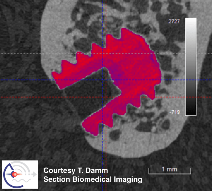
Parametric imaging is my area of expertise and I have pursued this in both preclinical and clinical investigations. In this webinar I hope I can relay to you that parametric imaging is fascinating because it can uncover information that is not visible for the naked eye. I will illustrate this with examples (e.g. imaging an implant in a mouse bone, see figure), and also present general concepts regarding the question: what defines the quality of quantitative data. Examples are taken from preclinical studies and I will outline strengths and limitations of a number of different imaging modalities. Hopefully, by listening to this webinar, you will be stimulated to use imaging approaches in your studies and will be better able to judge the quality of published quantitative imaging data.
Imaging can be done in 2D, 3D and 4D – for the latter I will highlight the challenges and benefits of sequential and time-lapse studies. After all, a video may be worth a million words!

 Download the app now and explore ECTS new mobile platform!
 iOS
iOS  Android